Showing posts with label Joint Pain. Show all posts

Wednesday, 7 August 2024

Posterior Pelvic Tilt

What is a Posterior Pelvic Tilt?

posterior pelvic tilt

A posterior rotation of the innominates is indicative of a posterior pelvic tilt. As a result, the person will typically appear to have a relatively flat back and "tucked under" buttocks. Even while the posterior tilt is less frequent than the anterior tilt, it still has many negative effects.

Because of the limited flexibility at the sacroiliac joints, the sacrum and lumbar spine move together with the pelvis when it moves. The vertebrae will stack more vertically on top of one another as a result of the posterior rotation, which will also lessen the lumbar lordosis. Reduction of compressive stresses in the spine and adequate shock absorption are two of the main purposes of lumbar lordosis.

Therefore, the spine's overall capacity to absorb shock is diminished when lumbar lordosis is lost. The intervertebral discs will experience greater compression stresses due to the vertebral bodies' more vertical placement on top of one another. Lumbar disc disease may be influenced by the intervertebral discs' increased degree of compression.

There are a number of causes of posterior pelvic rotations, but the majority result from long-term overuse of one's posture, such as slouching while sitting. If inadequate body mechanics, such as slouching, are repeatedly reinforced, it could worsen posterior pelvic rotation. Therefore, training good body mechanics and postural re-education are necessary in addition to short-term Physical therapies.

Causes of Posterior Pelvic Tilt

The posterior pelvic tilt may be caused by a number of muscle imbalance related causes. The pelvis may rotate posteriorly due to stiffness in the hamstrings and/or abdominal muscles. But because the hamstrings and abdominals are also phasic muscles, as they get tired, they lean toward weakening rather than hypertonicity.

Thus, this postural distortion cannot be caused by hamstring or abdominal tightness alone; rather, it requires a considerable degree of both. More often than not, posterior rotation is an adaptive habit continued by bad sitting and standing Body mechanics.

Posterior Pelvic Tilt Symptoms

Your lower back is under a lot of strain when you have a posterior pelvic tilt. Sciatica, or pain running down the back of one's thigh or glute, is one type of pain that might eventually result from this.

  • Weak and tight leg muscles
  • Shortened tendons around the pelvic bones
  • Feeling of tightness in abdominal muscles
  • Hamstring tightness
  • Improper balance
  • Poor posture
  • Reduced Lumbar Lordosis
  • Lower Back pain with or without Radiating pain in lower limb
  • Sciatica Pain
  • Hip Pain with or without Knee pain

A posterior pelvic tilt can be present by any of these variables. This is the moment when your upper body rounds back and your glutes tuck inward.

Long term Side Effects of Posterior Pelvic Tilt

Your entire posture is impacted because your neck and upper back are forced forward in an attempt to correct the improper pelvic position.

A forward head position and a more rounded shoulder, often known as thoracic kyphosis, are common symptoms of posterior pelvic tilt.

The compression strain on the mid and lower back will gradually rise due to posterior tilt. Due to the uneven loading of the spine caused by this increased compression stress, problems such as disk prolapse or disk herniation may result.

In addition, the lumbar curve's rounding places undue strain on your spine and raises the risk of hip, back, and leg pain.

Treatment of the Posterior Pelvic Tilt

The hamstring and abdominal muscle groups should be treated while treating posterior pelvic rotations. They require to get their tightness and myofascial trigger point presence looked upon. On the rectus abdominis muscle, longitudinal stripping and sweeping cross-fiber techniques can be used. It is possible to address the hamstring muscles' role in posterior pelvic tilting.

Treatment for the posterior pelvic tilt must include postural re-education, as was previously described. If soft tissue manipulation is tried without any kind of reinforced and corrected movement patterns, the practitioner is likely to have little success.

Stretches to Fix Pelvic Posterior Tilt:

Stretching Your Hamstrings While Seated

Long periods of sitting and standing can cause hamstring tightness, which can result in bad postures such a posterior pelvic tilt. Your hamstring will lengthen as a result of stretching, which will help your body maintain a more neutral pelvic position.

How to do it:

Sit down on a firm chair at first - Extend your left leg in front of you

Aim for three repetitions on each side. Bend forward and reach for your toes until you feel a minor stretch. 

Hold nearby for 10 to 20 seconds. Slowly return to the starting position. Repeat on your right side.

Take caution: If you extend the stretch too far, you risk pulling a muscle in your back. Take care not to go overboard. Try transferring this exercise on the floor if sitting in a chair is too challenging for you.

Bhujangasana: Abdominal Press Up (Cobra Pose)

Your tightened abdominal muscles will extend and become more flexible with the help of this stretch, which will enable a more neutral pelvic position.

How to carry it out:

- Start by lying on the floor with your hands flat at shoulder level. - Gradually push your hands up to lift your shoulder off the floor until you feel a slight stretch.

Exhale as you push deeper into the lumbar extension to increase your range of motion.  

Try to complete five repetitions.

Piriformis Stretch

The piriformis muscle may tighten up after extended periods of sitting. Stretching this muscle will enable the hip to move normally, which aids in achieving a neutral pelvic position.

How to do it:

Start by lying on your back with your feet flat on the ground and your knees bent.

To begin, place your right ankle over your left knee. Then, grab your left knee and draw it in towards your chest for a few seconds, until your right gluteal muscles stretch. Repeat on the other side. Try to complete three repetitions on each side.

Exercises to strengthen Muscles of the posterior pelvic tilt

Lunges:

Lunges improve quadriceps strength. It may be possible to correct a posterior pelvic tilt by strengthening these muscles.

How to carry it out:

- Start by placing your feet together. Stretch out your left leg in front of you. When your left leg is bowed to a 90-degree angle, your right knee should come into contact with the floor. Resuming your initial position requires pushing up with your left leg. On your right side, repeat. Try to complete three sets of ten lunges on each side.

* When in the lunge position, make sure your knee does not go over your toe as this puts more strain on the knee.

Superman:

Superman assists with correcting improper pelvic alignment by strengthening your lower back and glutes, which are connected to your pelvis.

The exercise can be performed as follows:

Lay on your stomach on the floor with your arms extended in front of you
Slowly raise your chest off the ground and attempt to hold this posture for 30 seconds
Slowly return your body to the beginning position
Try to complete three repetitions.

Leg Lifts

Leg raises assist in strengthening your hip flexors, which become incredibly weak due to the pelvic posterior tilt.

How to carry it out:

- In Supine Position on the soft mat with your legs extended. Keeping your arms straight by your sides, slowly raise your legs off the ground by contracting your abdominal muscles.

– Return your legs to the beginning position slowly.

The secret to this exercise is to solely move your legs; this will help your core muscles grow.

This is a challenging workout; if you have trouble lifting your legs, consider bending your knees a little.

Adjustments to Sleeping

To help you sleep with appropriate posture:

  • Stay away of sleeping on your stomach.
  • Place a little pillow below your knees if you prefer to sleep on your back.
  • A pillow should be placed between your knees if you are a side sleeper.
  • Put a tiny pillow or rolled towel beneath your back's arch if you sleep on your back.
  • Verify that the cushion and mattress suit the natural curve of your spine.

Correcting Posterior Pelvic Tilt While Sitting

Whether you sit all day at work or just a lot of it, it's important to make sure that:

  • You are sat in a posture-supporting chair.
  • Your choice of workstation or table should enable you to sit upright and prevent slouching.
  • You can either utilise an integrated lumbar support chair or use a pillow for this type of assistance.
  • You make the effort to move around and stretch, especially if you spend a lot of time seated.
Issues related to posterior pelvic tilt:

A posterior pelvic tilt puts more pressure on the lower back bones. This pressure can result in fatigue of the muscles as well as tension in the neck muscles.
  • Soreness in the lower back.
  • Rotation inward at the hip and knee.
  • Knee ache and hip pain.
  • Hip flexor tension is the cause of sciatica.

Summary

The tilting of the pelvis backward is called a posterior pelvic tilt. It mostly results from an imbalance in the muscles of the legs and the core, which has an impact on your body, activities, everyday posture, and work habits.

The symptoms can range in intensity and include low back pain, stiffness in the hamstring and abdominal muscles, and slouched posture, among others. Exercises that stretch and strengthen tense and weak muscles are the mainstay of treatment, along with changes to sitting and sleeping positions and other bad work habits.

Wednesday, 29 March 2023

Pes Anserine Bursitis: Cause, Symptoms, Treatment, Exercise


Pes Anserine Bursitis


What is a Pes Anserine Bursitis?

Pes anserine bursitis is a condition in which the bursa located at the inner aspect of the knee joint becomes inflamed. The pes anserine bursa is a small, fluid-filled sac that provides cushioning and reduces friction between the tendons of the hamstring muscles and the tibia bone of the lower leg.

This condition typically causes pain, tenderness, and swelling on the inside of the knee, just below the joint line. It is commonly seen in middle-aged women and people who are overweight or have osteoarthritis of the knee.

The most common causes of pes anserine bursitis include overuse or repetitive strain on the knee joint, injury, and degenerative changes in the joint due to aging or arthritis. Treatment options for pes anserine bursitis may include rest, ice, compression, elevation, physical therapy, anti-inflammatory medications, and in some cases, steroid injections or surgery.

Related Anatomy

The pes anserine bursa is located at the medial aspect of the knee, just below the joint line. It is situated between the tendons of three muscles:

  • Sartorius: a long, thin muscle that runs from the hip to the medial side of the tibia bone. It helps to flex and rotate the hip and knee joints.
  • Gracilis: a long, thin muscle that runs from the pubic bone to the tibia bone on the medial side of the knee joint. It helps to adduct and rotate the hip and flex the knee joint.
  • Semitendinosus: a long, thin muscle that runs from the ischial tuberosity of the pelvis to the tibia bone on the medial side of the knee joint. It helps to extend the hip and flex and medially rotate the knee joint.

The pes anserine bursa lies between these tendons and the underlying tibia bone. It helps to reduce friction between the tendons and the bone during movement.

Causes of Pes Anserine Bursitis

Pes anserine bursitis can be caused by a variety of factors, including:

  • Overuse or repetitive strain: Repeated stress on the knee joint can cause irritation and inflammation of the bursa, leading to pes anserine bursitis. This can occur in athletes who engage in activities that require a lot of running, jumping, or kicking.
  • Injury: Trauma to the knee, such as a direct blow or fall, can cause inflammation of the bursa.
  • Obesity: People who are overweight or obese are at a higher risk of developing pes anserine bursitis because the excess weight places more stress on the knee joint.
  • Osteoarthritis: This is a degenerative joint disease that can cause inflammation and damage to the knee joint, leading to pes anserine bursitis.
  • Other medical conditions: Some medical conditions such as gout, rheumatoid arthritis, or diabetes can increase the risk of developing pes anserine bursitis.
  • Improper form or training: Poor running or exercise technique, as well as inadequate training or warm-up, can put excessive stress on the knee joint and increase the risk of pes anserine bursitis.

Muscle imbalances: Weakness or imbalances in the hip or thigh muscles can cause abnormal stresses on the knee joint, leading to pes anserine bursitis.

Symptoms of Pes Anserine Bursitis

The most common symptoms of pes anserine bursitis include:

  • Pain: The pain is usually felt on the inside of the knee, just below the joint line. The pain may be sharp, dull, or throbbing, and may worsen with activity.
  • Swelling: The affected area may appear swollen and tender to the touch.
  • Stiffness: The knee may feel stiff or difficult to move, especially in the morning or after prolonged periods of inactivity.
  • Weakness: Weakness in the muscles around the knee, especially the quadriceps and hamstrings, may also be present.
  • Clicking or popping: Some people may experience clicking or popping sensations in the knee joint.
  • Limited range of motion: The range of motion in the knee joint may be limited, and it may be difficult to fully bend or straighten the knee.
  • Warmth or redness: In some cases, the skin over the affected area may feel warm or appear red.

It's important to note that these symptoms can also be caused by other knee conditions, so it's important to see a healthcare provider for an accurate diagnosis.

Risk factor

Several factors can increase the risk of developing pes anserine bursitis, including:

  • Age: Pes anserine bursitis is more common in middle-aged and older adults.
  • Gender: Women are more likely than men to develop pes anserine bursitis.
  • Obesity: Being overweight or obese increases the stress on the knee joint, which can lead to pes anserine bursitis.
  • Medical conditions: Certain medical conditions, such as osteoarthritis, rheumatoid arthritis, gout, and diabetes, can increase the risk of developing pes anserine bursitis.
  • Improper exercise technique: Poor running or exercise technique can put excessive stress on the knee joint, leading to pes anserine bursitis.
  • Muscle imbalances: Weakness or imbalances in the hip or thigh muscles can cause abnormal stresses on the knee joint, increasing the risk of pes anserine bursitis.
  • Sports and activities: Certain sports and activities that involve repetitive knee movements, such as running, cycling, or dancing, can increase the risk of pes anserine bursitis.
  • Previous knee injury: A previous knee injury, such as a ligament tear or fracture, can increase the risk of developing pes anserine bursitis.

Differential Diagnosis

There are several conditions that can cause similar symptoms to pes anserine bursitis, so it's important to have a healthcare provider perform a thorough examination to determine the correct diagnosis. Some possible differential diagnoses for pes anserine bursitis include:

  • Knee osteoarthritis: This is a degenerative joint disease that can cause pain, stiffness, and swelling in the knee joint.
  • Medial collateral ligament (MCL) injury: An injury to the MCL can cause pain, swelling, and instability in the knee joint.
  • Patellofemoral pain syndrome: This is a condition that causes pain in the front of the knee, often worsened by activities such as running or going up and down stairs.
  • Meniscal tear: A tear in the meniscus, the cartilage that cushions the knee joint, can cause pain, swelling, and difficulty with movement.
  • Osgood-Schlatter disease: This is a condition that typically affects adolescents and causes pain and swelling in the knee, especially during physical activity.
  • Rheumatoid arthritis: This is an autoimmune disease that can cause joint pain, swelling, and stiffness throughout the body, including the knee joint.
  • Gout of the Knee: This is a type of arthritis that occurs when uric acid crystals build up in the joints, causing pain, swelling, and inflammation.
  • Infection: In rare cases, an infection in the knee joint can cause symptoms similar to those of pes anserine bursitis.

Diagnosis

The diagnosis of pes anserine bursitis typically involves a physical exam, medical history, and diagnostic tests. During the physical exam, the healthcare provider may check for tenderness, swelling, and range of motion in the knee joint. They may also assess the strength of the muscles around the knee.

A medical history can help identify any potential risk factors, such as recent knee injury or medical conditions that may increase the risk of developing pes anserine bursitis.

Diagnostic tests may include imaging studies, such as X-rays or magnetic resonance imaging (MRI), to rule out other knee conditions such as osteoarthritis or a meniscal tear. Blood tests may also be ordered to check for signs of infection or underlying medical conditions.

In some cases, the healthcare provider may also perform a bursa aspiration, which involves using a needle to remove fluid from the affected bursa for further examination. This can help confirm the diagnosis of pes anserine bursitis and rule out other conditions that may cause similar symptoms.

Examination test for Pes Anserine Bursitis

During a physical examination, a healthcare provider may perform several tests to help diagnose pes anserine bursitis. Some of these tests include:

  • Palpation: The healthcare provider may press on the inside of the knee to check for tenderness and swelling in the pes anserine bursa.
  • Passive knee flexion: The healthcare provider may flex the knee while the patient is lying on their back, which can help identify any pain or discomfort in the pes anserine bursa.
  • Active resisted knee flexion: The healthcare provider may ask the patient to flex their knee against resistance, which can help identify any weakness in the muscles around the knee joint.
  • Ober's test: This test is used to assess the tightness of the iliotibial band, which can contribute to pes anserine bursitis. The patient lies on their side with their lower leg extended and their upper leg flexed at the hip and knee. The healthcare provider then lifts the upper leg and allows it to drop back down while keeping the hip extended. If the leg stays abducted (i.e., away from the midline of the body), it may indicate tightness in the iliotibial band.

Treatment of Pes Anserine Bursitis

Treatment for pes anserine bursitis typically involves a combination of self-care measures, physical therapy, and medication. In rare cases, surgery may be required.

  • Self-care measures: Resting the knee, applying ice packs to the affected area, and using over-the-counter pain relievers, such as ibuprofen or acetaminophen, can help relieve pain and reduce inflammation.
  • Physical therapy: Strengthening and stretching exercises can help improve the strength and flexibility of the muscles around the knee joint, which can help reduce stress on the bursa. The physical therapist may also use modalities such as ultrasound, heat or ice therapy to help reduce pain and swelling.
  • Medication: If self-care measures and physical therapy are not enough, the healthcare provider may prescribe stronger pain medications or corticosteroid injections to help reduce inflammation and relieve pain.
  • Surgery: In rare cases, surgery may be required to remove the affected bursa.
  • Preventative measures such as proper warm-up and cool-down exercises before and after physical activity, maintaining a healthy weight, and using proper technique during exercise can help prevent pes anserine bursitis from developing or recurring.

Physiotherapy treatment

Physiotherapy can be an effective treatment for pes anserine bursitis. A physical therapist can design a specific treatment plan based on the individual's symptoms, medical history, and physical examination.

  • Range of motion exercises: These exercises help improve the flexibility of the knee joint and surrounding muscles.
  • Strengthening exercises: Strengthening the muscles around the knee joint, such as the quadriceps, hamstrings, and glutes, can help reduce stress on the bursa and improve knee stability.
  • Manual therapy: Manual therapy techniques, such as massage or joint mobilization, can help improve joint mobility and reduce pain.
  • Modalities: Physical therapists can use modalities such as heat or ice therapy, ultrasound, and electrical stimulation to reduce pain and inflammation.
  • Gait training: Analyzing and correcting an individual's gait pattern can help reduce stress on the knee joint, which can reduce the risk of bursitis.
  • Education: Education on proper exercise techniques, posture, and footwear can help prevent bursitis from recurring.

It's important to work with a qualified physical therapist who can design a customized treatment plan based on the individual's specific needs and goals.

Exercises

Here are some examples of exercises that may be recommended by a physical therapist for pes anserine bursitis:

  • Quad sets: Sit on the floor with your legs straight out in front of you. Tighten the muscles at the front of your thigh, and press the back of your knee into the floor. Hold for 5 seconds, and then relax. Repeat for 10 repetitions, and gradually increase to 3 sets of 10 repetitions.
  • Hamstring stretches: Lie on your back with one leg extended straight out in front of you and the other bent with your foot flat on the ground. Hold the back of your thigh with both hands and gently pull your leg towards your chest until you feel a stretch in the back of your thigh. Hold for 30 seconds, and then repeat on the other leg. Repeat for 3-5 repetitions on each leg.
  • Clamshells: Lie on your side with your legs bent at a 90-degree angle. Keeping your feet together, lift your top knee as high as you can without moving your pelvis. Hold for 2 seconds, and then lower your leg back down. Repeat for 10 repetitions, and gradually increase to 3 sets of 10 repetitions.
  • Straight leg raises: Lie on your back with one leg bent and the other straight out in front of you. Tighten the muscles at the front of your thigh, and lift your leg up off the ground until it is parallel with your bent knee. Hold for 2 seconds, and then lower your leg back down. Repeat for 10 repetitions, and gradually increase to 3 sets of 10 repetitions.
  • Wall squats: Stand with your back against a wall and your feet shoulder-width apart. Slide down the wall until your knees are bent at a 90-degree angle. Hold for 10-30 seconds, and then slide back up. Repeat for 10 repetitions, and gradually increase to 3 sets of 10 repetitions.

It's important to only perform these exercises under the guidance of a qualified physical therapist to ensure proper technique and to avoid exacerbating symptoms.

How to Prevent Pes Anserine Bursitis?

Here are some tips to help prevent pes anserine bursitis:

  • Warm-up and cool-down: Always properly warm-up before exercising, and cool-down afterwards. This helps prepare the muscles for activity and reduces the risk of injury.
  • Strengthen and stretch: Strengthen the muscles around the knee joint, such as the quadriceps, hamstrings, and glutes, and stretch these muscles regularly to improve flexibility and reduce the risk of injury.
  • Wear proper footwear: Choose appropriate shoes for your physical activities, such as running shoes for running or cross-training shoes for weight lifting.
  • Maintain a healthy weight: Excess weight can put additional stress on the knees and increase the risk of injury.
  • Use proper technique: Make sure to use proper technique during exercises to avoid putting unnecessary stress on the knees.
  • Gradually increase activity level: Gradually increase the intensity, duration, and frequency of physical activity to avoid overuse injuries.
  • Avoid repetitive activities: Try to vary your physical activities to avoid repetitive stress on the knee joint.

By following these tips, you can help reduce your risk of developing pes anserine bursitis and other knee injuries.

Pes Anserine Bursitis recovery time

The recovery time for pes anserine bursitis can vary depending on the severity of the injury and the individual's response to treatment. In mild cases, recovery may only take a few weeks, while more severe cases may take several months to fully heal.

It's important to allow enough time for the bursa to heal completely before returning to physical activity. Continuing to exercise or play sports too soon can prolong the healing process and increase the risk of reinjury.

Working with a qualified healthcare provider, such as a physical therapist or orthopedic specialist, can help ensure an appropriate treatment plan is developed and followed for optimal recovery. Adhering to the treatment plan, including rest, physical therapy, and other recommended interventions, can help speed up recovery time and reduce the risk of recurrence.

Summary

Pes anserine bursitis is a condition characterized by inflammation of the bursa located between the tendons of the hamstring muscles and the shinbone. It typically causes pain and swelling on the inside of the knee and can be caused by overuse, injury, or underlying medical conditions.

Risk factors for pes anserine bursitis include obesity, previous knee injuries, and conditions such as osteoarthritis or diabetes. Diagnosis typically involves a physical exam, imaging tests, and ruling out other potential causes of knee pain.

Treatment for pes anserine bursitis includes rest, ice, compression, and elevation (RICE), medications, physical therapy, and in some cases, corticosteroid injections or surgery. Physical therapy can help improve strength and flexibility in the muscles surrounding the knee joint, and specific exercises such as quad sets, hamstring stretches, clamshells, straight leg raises, and wall squats may be recommended.

Preventive measures include warming up and cooling down before and after exercise, wearing appropriate footwear, maintaining a healthy weight, using proper technique, and gradually increasing activity levels. Recovery time can vary depending on the severity of the injury and the individual's response to treatment, but it's important to allow enough time for the bursa to heal completely before returning to physical activity.

Tuesday, 28 March 2023

Shoulder Impingement Syndrome: Cause, Symptoms, Treatment, Exercise

What is a Shoulder impingement syndrome?

Shoulder Impingement Syndrome

Shoulder impingement syndrome (SIS) is a condition where the soft tissues in the shoulder joint become compressed or pinched, causing pain, inflammation, and limited movement. This compression typically occurs in the subacromial space, which is the narrow passageway between the acromion (a bony protrusion at the top of the shoulder blade) and the rotator cuff tendons.

The rotator cuff is a group of four muscles and their tendons that surround the shoulder joint and help to stabilize and move the shoulder. When the rotator cuff tendons become inflamed or irritated, they can swell and thicken, making it more difficult for them to move smoothly through the subacromial space. This can lead to rubbing and compression of the tendons and other soft tissues, causing pain and discomfort.

SIS can be caused by a variety of factors, including overuse, poor posture, muscle imbalances, and structural abnormalities in the shoulder joint. It is a common condition among athletes, particularly those who engage in overhead activities such as throwing, swimming, or weightlifting.

Treatment for SIS typically involves rest, physical therapy, and anti-inflammatory medications. In more severe cases, surgery may be necessary to relieve compression in the subacromial space.

Related Anatomy

To understand shoulder impingement syndrome (SIS), it is important to have a basic understanding of the anatomy of the shoulder joint.

The shoulder joint is a ball-and-socket joint that connects the upper arm bone (humerus) to the shoulder blade (scapula). The ball of the joint is formed by the rounded end of the humerus, while the socket is formed by a shallow depression on the scapula called the glenoid fossa. The joint is stabilized by a group of muscles and tendons called the rotator cuff, which wraps around the head of the humerus and helps to hold it in place within the socket.

The subacromial space is a narrow passageway between the acromion, a bony projection of the scapula, and the rotator cuff tendons. The space is lined with a lubricating sac called the subacromial bursa, which helps to reduce friction as the tendons move through the space. When the rotator cuff tendons become inflamed or irritated, they can swell and thicken, making it more difficult for them to move smoothly through the subacromial space. This can lead to rubbing and compression of the tendons and other soft tissues, causing pain and discomfort.

Other important structures in the shoulder joint include the labrum, a ring of cartilage that helps to deepen the socket and provide additional stability to the joint, and the biceps tendon, which attaches the biceps muscle to the shoulder blade and helps to stabilize the joint during certain movements.

What are the causes of Shoulder impingement syndrome?

Shoulder impingement syndrome (SIS) can be caused by a variety of factors, including:

  • Overuse: Activities that involve repetitive overhead movements, such as throwing a ball or painting a ceiling, can put a lot of stress on the rotator cuff tendons and lead to inflammation and irritation.
  • Poor posture: Poor posture can cause the shoulder joint to become misaligned, which can put extra pressure on the rotator cuff tendons and lead to impingement.
  • Muscle imbalances: Imbalances in the muscles that surround the shoulder joint can cause the joint to become unstable and increase the risk of impingement.
  • Structural abnormalities: Some people are born with structural abnormalities in the shoulder joint, such as a hooked acromion or a narrow subacromial space, which can increase the risk of impingement.
  • Trauma: A fall or other injury that causes damage to the rotator cuff tendons or other soft tissues in the shoulder can lead to impingement.
  • Aging: As we age, the tendons in the rotator cuff can become weaker and more prone to injury and inflammation, which can increase the risk of impingement.

Other conditions: Certain medical conditions, such as arthritis, can cause inflammation and swelling in the shoulder joint, which can lead to impingement.

Symptoms of Shoulder impingement syndrome

The symptoms of shoulder impingement syndrome (SIS) can vary depending on the severity of the condition, but common symptoms include:

  • Pain: Pain in the shoulder joint is the most common symptom of SIS. The pain is usually felt in the front or side of the shoulder and may be sharp or dull. The pain may be worse when the arm is lifted overhead or when the arm is moved away from the body.
  • Weakness: Weakness in the affected arm is another common symptom of SIS. This can make it difficult to perform certain activities that require lifting or reaching, such as putting on a shirt or reaching for a high shelf.
  • Limited range of motion: Impingement can limit the range of motion in the shoulder joint, making it difficult to move the arm in certain directions.
  • Clicking or popping sounds: Some people with SIS may experience clicking or popping sounds in the shoulder joint when they move their arm.
  • Swelling or inflammation: Inflammation and swelling in the shoulder joint may be present in more severe cases of SIS.
  • Pain at night: Some people with SIS may experience pain that wakes them up at night, particularly if they sleep on the affected side.

If you are experiencing any of these symptoms, it is important to see a healthcare professional for an accurate diagnosis and appropriate treatment.

Risk factor

There are several risk factors that can increase the likelihood of developing shoulder impingement syndrome (SIS), including:

  • Age: SIS is more common in older adults, as the tendons in the rotator cuff can weaken and become more prone to injury and inflammation with age.
  • Overuse: Repetitive overhead activities, such as throwing a ball or painting a ceiling, can increase the risk of SIS by putting a lot of stress on the rotator cuff tendons.
  • Poor posture: Poor posture can cause the shoulder joint to become misaligned, which can increase the risk of impingement.
  • Muscle imbalances: Imbalances in the muscles that surround the shoulder joint can cause the joint to become unstable and increase the risk of impingement.
  • Structural abnormalities: Some people are born with structural abnormalities in the shoulder joint, such as a hooked acromion or a narrow subacromial space, which can increase the risk of impingement.
  • Previous shoulder injuries: Previous injuries to the shoulder, such as a dislocated shoulder or a rotator cuff tear, can increase the risk of developing SIS.
  • Certain sports or activities: Athletes who engage in sports that require a lot of overhead movements, such as baseball or swimming, are at an increased risk of developing SIS.
  • Occupation: Jobs that require repetitive overhead activities, such as construction or painting, can increase the risk of developing SIS.
  • Genetics: Some people may be more predisposed to developing SIS due to genetic factors that affect the structure and function of the shoulder joint.

It's important to note that having one or more of these risk factors doesn't necessarily mean that someone will develop SIS, but it does increase their likelihood of developing the condition.

Differential Diagnosis

Shoulder impingement syndrome (SIS) can have similar symptoms to other conditions affecting the shoulder, so it's important to consider differential diagnoses when evaluating a patient with shoulder pain. Some potential differential diagnoses for SIS include:

  • Rotator cuff tear: A rotator cuff tear is a tear in one or more of the rotator cuff tendons that can cause pain, weakness, and limited range of motion in the shoulder joint.
  • Bursitis: Bursitis is inflammation of the bursae, small fluid-filled sacs that cushion the joints. In the shoulder, bursitis can cause pain, swelling, and limited range of motion.
  • Frozen shoulder: Frozen shoulder, also known as adhesive capsulitis, is a condition in which the shoulder joint becomes stiff and painful, often as a result of injury or surgery.
  • Shoulder arthritis: Arthritis in the shoulder joint can cause pain, stiffness, and limited range of motion in the shoulder.
  • Labral tear: A labral tear is a tear in the labrum, a ring of cartilage that surrounds the shoulder joint. This can cause pain, weakness, and limited range of motion in the shoulder.
  • Cervical spine disorders: Disorders affecting the cervical spine, such as cervical radiculopathy or herniated disc, can cause pain that radiates into the shoulder.
  • Thoracic outlet syndrome: Thoracic outlet syndrome is a condition in which the nerves and blood vessels that pass through the thoracic outlet, a narrow space between the collarbone and first rib, become compressed. This can cause pain and numbness in the shoulder and arm.

A thorough evaluation by a healthcare professional can help differentiate between these conditions and determine the most appropriate treatment plan.

Diagnosis

The diagnosis of shoulder impingement syndrome (SIS) is typically made by a healthcare professional, such as a physician, orthopedist, or physical therapist. The diagnostic process usually involves a combination of a physical examination, medical history, and imaging tests.

During the physical examination, the healthcare professional will evaluate the range of motion of the shoulder joint, assess for areas of tenderness or swelling, and test the strength of the rotator cuff muscles. They may also perform special tests, such as the Neer or Hawkins-Kennedy test, which can help confirm a diagnosis of SIS.

A medical history will also be taken to assess for risk factors and previous injuries that may have contributed to the development of SIS.

Imaging tests, such as X-rays, ultrasound, or magnetic resonance imaging (MRI), may be ordered to confirm the diagnosis and evaluate the severity of the condition. X-rays can help rule out other conditions, such as arthritis or a fracture, while ultrasound or MRI can provide more detailed images of the soft tissues in the shoulder joint, such as the rotator cuff tendons and bursae.

Overall, a combination of physical examination, medical history, and imaging tests can help healthcare professionals diagnose SIS and develop an appropriate treatment plan.

Treatment of Shoulder impingement syndrome

The treatment of shoulder impingement syndrome (SIS) depends on the severity of the condition, as well as the patient's individual needs and goals. In general, treatment for SIS may include the following:

  • Rest and activity modification: Resting the affected shoulder and avoiding activities that exacerbate the symptoms can help reduce pain and inflammation in the shoulder joint.
  • Physical therapy: Physical therapy exercises can help improve the strength and flexibility of the rotator cuff muscles and improve the stability of the shoulder joint. This can help reduce the risk of impingement and improve shoulder function.
  • Non-steroidal anti-inflammatory drugs (NSAIDs): Over-the-counter pain medications, such as ibuprofen or naproxen, can help reduce pain and inflammation in the shoulder joint.
  • Corticosteroid injections: In some cases, corticosteroid injections may be used to reduce inflammation in the shoulder joint and provide pain relief.
  • Surgery: In severe cases of SIS, surgery may be necessary to remove bone spurs or repair tears in the rotator cuff tendons. Surgery may also be recommended if conservative treatments have not been effective.
  • Lifestyle modifications: Making lifestyle modifications, such as improving posture, maintaining a healthy weight, and avoiding activities that place excessive stress on the shoulder joint, can help reduce the risk of SIS and improve shoulder health.

It's important for patients with SIS to work closely with their healthcare professionals to develop a personalized treatment plan that addresses their specific needs and goals. With appropriate treatment, most people with SIS can expect to see improvements in their symptoms and shoulder function.

Physiotherapy treatment

Physiotherapy is a common and effective treatment option for shoulder impingement syndrome (SIS). Physiotherapy treatment for SIS may include the following:

  • Range of motion exercises: Gentle range of motion exercises can help improve the flexibility of the shoulder joint and reduce stiffness.
  • Strengthening exercises: Specific exercises that target the rotator cuff muscles can help improve strength and stability in the shoulder joint. This can help reduce the risk of impingement and improve shoulder function.
  • Posture training: Poor posture can contribute to the development of SIS. Physiotherapists can provide guidance on proper posture and teach patients how to maintain good posture throughout the day.
  • Manual therapy: Manual therapy techniques, such as massage or joint mobilization, can help reduce pain and improve mobility in the shoulder joint.
  • Modalities: Modalities, such as ice or heat therapy, ultrasound, or electrical stimulation, can help reduce pain and inflammation in the shoulder joint.
  • Education: Physiotherapists can provide education on proper body mechanics and ergonomics to reduce stress on the shoulder joint during daily activities.

Home exercise program: Physiotherapists can develop a customized home exercise program for patients to continue their exercises outside of the clinic and improve their shoulder function.

Overall, physiotherapy can be a highly effective treatment option for SIS. Working with a qualified physiotherapist can help patients reduce their pain, improve their shoulder function, and prevent the condition from worsening.

Exercise for Shoulder impingement syndrome

Exercise is an important component of treatment for shoulder impingement syndrome (SIS). However, the type and intensity of exercises will depend on the severity of the condition and the patient's individual needs and goals. Here are some exercises that may be helpful for patients with SIS:

  • Pendulum exercise: Stand with one hand resting on a table or chair. Swing the opposite arm gently back and forth, and then in circles. This exercise can help improve range of motion and reduce stiffness in the shoulder joint.
  • Scapular stabilization exercises: These exercises are designed to improve the strength and stability of the muscles surrounding the shoulder blade, which can help reduce the risk of impingement. Examples of scapular stabilization exercises include scapular retractions, wall slides, and shoulder blade squeezes.
  • Rotator cuff strengthening exercises: Strengthening the rotator cuff muscles can help improve shoulder stability and reduce the risk of impingement. Examples of rotator cuff strengthening exercises include external rotations, internal rotations, and prone shoulder flexion.
  • Shoulder stretches: Stretching the muscles in the shoulder can help improve flexibility and reduce tension in the joint. Examples of shoulder stretches include cross-body shoulder stretch, chest stretch, and shoulder blade stretch.
  • Resistance band exercises: Resistance band exercises can be used to strengthen the shoulder muscles and improve shoulder function. Examples of resistance band exercises include external rotation with resistance band, internal rotation with resistance band, and shoulder abduction with resistance band.

It's important for patients with SIS to work closely with a qualified healthcare professional, such as a physiotherapist or exercise physiologist, to develop a personalized exercise program that addresses their specific needs and goals. Patients should also listen to their body and stop any exercise that causes pain or discomfort.

Sunday, 28 August 2022

Heel Pain Cause, Symptoms, Diagnosis, Treatment, Exercise

Heel Pain
Heel Pain

If you're experiencing heel pain, you're not alone. Heel pain is one of the most common types of foot pain, and it can be caused by a variety of factors. In this blog post, we'll discuss the most common causes of heel pain, Symptoms, Diagnosis and provide tips for how to treat it.

Causes of Heel Pain

Heel pain can have many different causes. It can be caused by an injury, by wearing shoes that don't fit properly, or by a medical condition.

If you have heel pain, it is important to see a doctor to find out the cause. Treating the cause of the pain will help you to get rid of the pain.

Wearing shoes that don't fit properly can also cause heel pain. Shoes that are too tight or too loose can rub against the heel, causing pain. Shoes that are too high can also put pressure on the heel.

Some medical conditions can also cause heel pain. One of the most common is plantar fasciitis, which is an inflammation of the tissue that connects the heel to the toes. Other conditions that can cause heel pain include arthritis, gout, and bone fractures.

Heel Spurs

One of the most common causes of heel pain is heel spurs. Heel spurs are bony growths that form on the heel bone, and they can cause pain and inflammation. Heel spurs are typically caused by repetitive stress on the heel, such as from running or jumping.

There are a few ways to treat heel spurs. In many cases, rest, ice, and elevation can help to relieve pain and inflammation. You may also need to wear a heel spur pad or heel cup to reduce stress on the heel. If conservative measures don't provide relief, you may need surgery to remove the heel spur.

Plantar Fasciitis

Another common cause of heel pain is plantar fasciitis. Plantar fasciitis is a condition that causes inflammation and pain in the plantar fascia, a band of soft tissue that runs along the bottom of the foot - that connects the heel to the toes.. Plantar fasciitis is most commonly caused by overuse or repetitive stress on the plantar fascia of the foot.

There are a few ways to treat plantar fasciitis. In many cases, rest, ice, and elevation can help to relieve pain and inflammation. You may also need to wear a heel spur pad or heel cup to reduce stress on the heel. If conservative measures don't provide relief, you may need surgery to release the plantar fascia.

If you are experiencing heel pain, it is important to see a doctor to determine the cause and get treatment. Heel pain can be a sign of a serious condition, such as plantar fasciitis or a heel spur, so it is important to get it checked out.

Other conditions that can cause heel pain include arthritis, gout, and bone fractures.

Symptoms:

The Heel pain can range from a dull ache to sharp, stabbing pain, and it can be mild or severe. If you’re experiencing heel pain, here are a few of the most common symptoms:

  • Pain in the heel or ankle, which may worsen when you walk or stand
  • Inflammation or swelling in the heel
  • Redness or warmth in the heel
  • Stiffness of the Ankle joint or in the heel
  • Difficulty walking or putting weight on the heel

If you’re experiencing any of these symptoms, it’s important to see a doctor to rule out any underlying medical conditions and get the proper diagnosis and treatment.

Diagnosis:

There are many different possible causes of heel pain, so a thorough diagnosis is important. Heel pain can be caused by structural problems with the foot, such as heel spurs or plantar fasciitis. It can also be caused by problems with the bones, muscles, or tendons in the foot or lower leg. In some cases, heel pain is the result of a nerve problem. Proper diagnosis of the Heel pain is highly important for treatment.

Your Doctor examine your foot and Heel, ask you few quetions regarding your pain and also prescribe you X-Ray or MRI (Magnatic Resonance Imaging) test.

Conservative treatments for heel pain include rest, ice, and over-the-counter anti-inflammatory medications. If these treatments don't provide relief, your doctor may recommend more aggressive measures, such as physical therapy, steroid injections, or last option is surgery.

Treatment of Heel Pain:

Heel pain is a common complaint that can be caused by a variety of conditions. Although most heel pain is manageable with conservative treatment, it is important to see a doctor to rule out serious underlying conditions.

RICE Principle
RICE Principle

There are a number of things that can be done to help relieve heel pain. Most people will need to see a doctor to get a diagnosis and follow treatment plan. 

Treatment options may include pain relieving medications, with use of ice pack and heat therapy, with few days rest.

If these treatments don't provide relief, your doctor may recommend more aggressive measures, such as physical therapy, steroid injections, or last option is surgery.

In some cases, surgery may be needed if Medical and Physical therapy treatment not help you to recover.

Most heel pain can be managed successfully with conservative treatment. However, if you're still experiencing pain after several weeks of self-care, be sure to see your doctor for a more thorough evaluation.

Exercise

If you are suffering from heel pain, there are certain exercises you can do to help lessen the pain. These exercises will help stretch and massage the muscles in your foot, and give relief to your Achilles tendon.

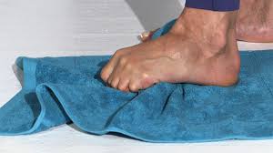
Towel Scrunches exercise

Towel Scrunches exercise


One exercise is to sit down and place a rolled up towel under your foot. While keeping your foot flexed, slowly roll the towel back and forth under your foot for a few minutes. This will help stretch and massage the plantar fascia in your foot.

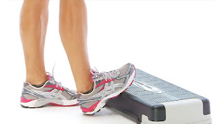
Calf Stretching Exercise


Calf Stretching Exercise

Another exercise you can do is to stand on a step and let your heel hang off the edge. Gently stretch your foot by moving your heel up and down. You should feel a stretch in your Achilles tendon. Hold this stretch for a few seconds and then release. Repeat a few times.


Ergonomics of Heel Pain:

Heel pain is a common issue that can be caused by a variety of factors. Poorly fitting shoes, overuse, and injury are all common causes of heel pain.

There are a few things you can do to help prevent heel pain. First, make sure you wear shoes that fit properly and support your feet. 

Secondly, avoid overdoing it when you are participating in activities that put stress on your feet and heels. If you do start to experience heel pain, be sure to rest and give your feet a break.

If you are suffering from heel pain, there are a few things you can do to find relief.  Pain relieving medication and oil can help to ease the pain. You can also try icing the affected area for 20 minutes at a time. If the pain is severe, you may need to see a doctor or physical therapist for more aggressive treatment.

Thursday, 25 August 2022

Knee Pain Cause, Symptoms, Diagnosis, Treatment & Exercise

Knee Pain Treatment
Knee Pain
Introduction

Knee pain is one of the most common complaints in the world. It occurs due to a number of reasons like overuse, injury or arthritis. There are many types of knee pain which can be classified into 4 major categories: 

1. Pain due to overuse:

Pain in the knee is caused by overuse of the joint which can be due to activities like running, jumping or climbing stairs. Overuse of the knee joint may lead to pain and swelling which usually goes away after a few days if it is mild and rest. 

2. Pain due to injury: 

Injury of the knee can occur when there is a sudden impact on the joint like falling down while playing or sports. In this case, there will be swelling and bleeding as well as bruising around the knee area which may last for 1-2 weeks even after proper medical treatment.

3. Pain due to arthritis: Arthritis is the inflammation of the joints which causes pain, swelling and stiffness in them. The condition can be acute or chronic depending on how long it lasts. 

4. Pain due to bursitis: Bursitis is another type of inflammation which can occur in the knee area as well. 

This condition occurs when there is friction between the bones, tendons or ligaments which causes swelling and pain around the joint.

Knee pain cause


Knee pain is a common problem, and it can be caused by many things.
In fact, knee pain isn’t just one thing! 
It can be caused by any number of things:
  • A twisted Knee joint, ankle or foot
  • A torn ligament or tendon in your knee (particularly the anterior cruciate ligament)
  • An arthritic joint (the ball and socket joint) A blow to the knee joint A muscle strain or sprain A condition called chondromalacia (which causes inflammation of the cartilage in your knee)
  • Pain and swelling around the knee joint A meniscus tear (a tear to the cartilage in the knee joint)
  •  A knee injury from playing sports (such as football, basketball and skiing) 
  • A knee injury that causes your kneecap to dislocate (also called a patellar subluxation)
  • A knee injury that damages the nerves in your knee joint A knee injury caused by an infection
  •  A knee injury that causes damage to the ligaments of your knee joint A knee injury caused by a fall  A knee injury caused by an accident (such as a car crash) A genetic condition called osteogenesis imperfecta (which causes brittle bones)
Knee pain symptoms

The most common complaint of knee pain is a burning sensation in the joint. Other symptoms include:
  • Pain in the knee joint when you bend, straighten, or flex the knee
  • Pain in the knee joint when walking or running.
  • Pain in the knee joint when climbing stairs (or going up and down them)
  • Pain in the knee joint while squatting or kneeling. 
  • Pain in the knee joint when lying on your back with your legs straight out.
  • Pain in the knee joint that goes away after exercise and then comes back later.
  • You may also experience swelling around your knee joint if you’ve injured it.

Torn meniscus symptoms include:  Pain in the knee joint when you bend, straighten, or flex the knee
You may also experience swelling around your knee joint if you’ve injured it.

Knee pain diagnosis

Knee pain diagnosis can be difficult. Your doctor will ask you about the symptoms you’re experiencing and what activities trigger them. They may also examine your knee joint to see if they can find a cause for the pain. If they suspect that you have a torn meniscus, they may order an MRI scan or x-ray to confirm this diagnosis. 

Knee pain treatment

Treatment for knee pain is a combination of rest, ice, compression and elevation (RICE). This treatment can be done at home by yourself or with the help of a physical therapist. The goal is to allow your body time to recover from the injury so that it doesn't worsen into something more serious later on.

Physical therapy is another important part of your treatment. A physical therapist can help you strengthen the muscles that support your knee, as well as improve flexibility and range of motion. They can also help you identify any biomechanical issues that may be causing pain in your knee.

If the pain is severe and long-lasting, your doctor may suggest that you see an orthopedic surgeon. This is because they have more experience of treating knee injuries than general practitioners or family doctors. 
The type of treatment you receive will depend on how severe your Knee pain or injury is.

 If you have a torn meniscus, your doctor will recommend few treatment options. You may be given pain relief medication to help manage the symptoms of your injury.

Use of RICE Principle to Relive Knee pain:

  • Resting and icing your knee as soon as possible.
  • Elevating your leg above heart level when resting or sleeping.
  • Using a knee immobilizer to keep the joint stable while it heals. 
  • Using crutches to help support your weight while walking. 
  • Wearing a brace that stabilizes the knee joint to prevent further injury.
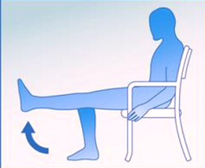
Knee pain exercise
Knee pain exercise
Knee pain exercise 


Knee pain exercises are important to strengthen the muscles around the knee. Knee pain exercises can help you to recover from knee pain and prevent it from occurring again in the future.

Knee pain exercises can also improve your condition and make it more comfortable, which can give you more energy as well as make moving easier, so there's no reason not to do them!

As well as being useful for preventing injury, knee pain exercises can also help to make your knee stronger and more flexible. This is important because it means that you are less likely to injure yourself again in the future.

Knee pain exercises can also be useful for helping to prevent other injuries in the future. For example, if you have knee pain and then start doing regular exercises, your knee will become stronger and more flexible over time. This means that it is less likely to happen again.

How to prevent Knee pain?

  • Modify your lifestyle. If you’re overweight or obese, losing weight can help reduce knee pain. 
  • Reduce the amount of alcohol you drink and stop smoking—both of these habits can increase your risk for osteoarthritis. 
  • Try to exercise regularly, even if it’s just walking around your neighborhood. Exercise strengthens muscles around joints, making them less likely to be injured during everyday activities.
  • Wear sports shoes with good support.
  • Limit the amount of time you spend sitting, especially in a low chair or on a couch.
  • Exercise regularly to strengthen your muscles and improve joint movement.
  • Consider changing jobs if you’re often on your feet all day at work.
Conclusion

Knee pain is a common problem among people. Knee pain can be caused by various reasons like arthritis, injury or even due to physical exertion. If you are suffering from knee pain and want to know more about it, then you must read this article thoroughly. 

We have discussed here about what causes knee pain in detail and also how can treat it effectively with some tips and tricks that might help you out with your condition.

Monday, 4 April 2022

Ankle pain without Injury: Cause, Symptoms, Treatment, Exercise

Ankle pain without Injury

If you have Sudden Ankle pain without injury, there may too many cause of this pain and symptoms are vary according to conditions. Treatment of Ankle pain is also depends upon cause, symptoms and diagnosis of Ankle pain.

In this article we discuss what are the possible cause of Ankle pain without Injury and symptoms. we also discuss how to diagnose ankle pain, Medical treatment and Physiotherapy exercise.

What are the causes of Ankle pain without injury?

There are too many causes of Ankle pain still if you are not injured, few most common cause of Ankle pain are:

  • Overuse of Ankle joint- continuous work load mainly farmers and laborers require 8 to 10 hours work in standing and walking position, these may lead to pressure on the joint and causes pain
  • osteoarthritis of Ankle joint: These are other cause of Ankle pain in which age related wear and tear of the Ankle joint may leads to damaged to joint surface.
  • Bursitis: Bursae (Fluid-filled sacs) arount the joint which provides cushion to your bones when they move. When bursae become irritated and inflamed called Bursitis.
  • Tendonitis: Muscle's tendon become irritated, inflamed called tendonitis. These tendons connect muscles to bones. Continuous workload may lead stress on the muscle's tendon such as Achilles tendinitis.
  • Plantar fasciitis causes pain in the bottom of the heel. The plantar fascia is a thin ligament that connects your heel to the front of your foot. It supports the arch in your foot and is important in helping you walk.
  • Flatfoot: When your arch is not properly develop from birth called congenital flatfoot which may lead to pain and swelling in the ankles and feet.
  • Gout: When uric acid increases in your body, causes multiple joint pain including Ankle joint. These uric acid created crystals which are deposited in the joint and causes joint pain, swelling and also restrict the movement most affect both the Ankles.

What are the signs and symptoms of ankle pain without injury?

  • Ankle pain with swelling
  • Bruising
  • Redness
  • Numbness or tingling
  • Instability
  • Ankle Joint Pain
  • Inability to bear weight on the affected ankle
  • Ankle joint Stiffness
  • Weakness
  • Discoloration
  • Warmth
  • Tenderness
  • Difficulty in walking
Diagnosis Ankle pain without Injury:

During a physical examination, the Physiotherapist will examine the patient’s ankle, foot, and lower limb. The doctor will also check the area of skin around the injury for proper points of tenderness and move the patient’s Ankle to check the range of motion and to know what positions cause discomfort or pain. 

Following are the imaging study also helpful for differential diagnosis of Ankle pain:

X-ray: X-Ray is mainly helpful if any arthritis changes are seen in the joint.
Magnetic resonance imaging (MRI): If x-ray is not helpful then further investigation Doctor recommend you MRI which uses radio waves & a strong magnetic field to produce detailed cross-sectional or 3-D images of the soft internal tissue of the ankle, with ligaments.
CT scan: If Doctor can get more detail view regarding the bones of the joint. CT scan takes too X-rays from many variant angles and mixes them to make cross-sectional or 3-D images.
Ultrasound: An ultrasound uses sound waves to produce images. These images may help doctors find the condition of a ligament/tendon when the foot is in a different position.

Treatment of Ankle pain without injury:

Medical treatment:

Mostly your Doctor prescribes you pain relieving medicine mostly NSAIDs are most common such as Diclofenac, Aceclefenac are enough to manage the ankle pain with Pain relieving gel and patches.

Home remedies for Ankle pain you can do is mainly use of Hot Packs- Cold Packs that helps to relieve pain and swelling naturally.

Physiotherapy treatment:

Physiotherapy treatment are mostly depends upon cause, symptoms and diagnosis of your Ankle pain. Most commoly Physiotherapy treatment in Ankle pain are pain relieving Electrotherapy modalities and Exercise.

Electrotherapy treatment:

There are various electro. modalities are uses to relive pain, swelling mainly in ankle pain three modalities would be beneficial which are describe below:

Ultrasound: Ultrasound is usually apply on tender point to relieve of pain.
TENS (Transcutaneous electrical nerve stimulation): TENS modality is mainly for reduction of pain, swelling.
IFT (Interferential Therapy ): If patient have any type of swelling, pain, inflammation ,IFT would be most effective modality.

Exercises for Ankle pain:

Regular exercise must help the patient to relieve pain and strengthen weak muscles.

Stretching exercise:

Achilles tendon & plantar fascia stretch:

To do this exercise, you can loop a towel around the ball of your foot & pull it back towards your body with a straight knee. Hold for 10 to 20 seconds for each foot and do it 3 times.

Sitting plantar fascia stretch:

To do this exercise you should cross your foot over the opposite knee. The base of the toes is put on the ground & gently pull back until you feel a stretch along the innerside of your foot. Hold for 10 to 30 seconds per repetition. Repeat 3 times on each foot.

Plantar fascia stretch:

To do this exercise you should put a towel on the floor in front of your chair, with the heels which are on the ground, try to pick up the towel by grabbing it with your toes, this exercise Repeat 10-15 times with 2 times a day.

Standing calf stretch:
Standing calf stretch

To do this exercise, you must stand facing a wall then place both hands on the wall which is in front of you. Put one foot forward to the other with the front foot about 30cm away from the wall, keep the other knee straight with keep your back heel flat on the floor. Gradually bend the front knee until you feel gentle stretch in the calf of the back lower leg. Relax and repeat 8 to 10 times. This exercise may also be done with the back knee slightly bent for a milder stretch.

Deep calf stretch:

To do this exercise you must stand on a step with the heel hanging off the edge. Your may also allow holding onto something for support. You should gradually drop heel towards the floor and feel a stretch in the calf muscles & Achilles tendon. Hold this for 10 to 30 seconds. Repeat this exercise 2-3 times.