Showing posts with label Ankle pain. Show all posts

Saturday, 15 April 2023

Peroneal tendon tear: Cause, Symptoms, Treatment, Exercise

What is a Peroneal tendon tear?

Peroneal tendon tear

Peroneal tendon tear is a condition where one or both tendons that run down the outer side of the lower leg, behind the ankle and into the foot, are partially or completely torn. These tendons are responsible for stabilizing the ankle and foot during movement, especially during activities that involve twisting or turning of the foot. 

A tear in the peroneal tendons can lead to pain, weakness, swelling, and instability in the ankle and foot. It can be caused by repetitive strain, overuse, or traumatic injury to the tendons. Treatment for peroneal tendon tear may involve rest, immobilization, physical therapy, or surgery depending on the severity of the injury.

Related Anatomy

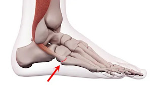
The peroneal tendons are two fibrous cords that originate from the muscles in the outer part of the lower leg, the peroneus longus, and peroneus brevis muscles. The peroneus longus tendon runs under the foot and attaches to the first metatarsal bone, while the peroneus brevis tendon runs behind the ankle and attaches to the fifth metatarsal bone. 

These tendons work together to stabilize the ankle and foot during movement, especially during activities that involve twisting or turning of the foot. The tendons are surrounded by a protective sheath and held in place by ligaments and other soft tissues. The blood supply to the peroneal tendons is limited, which can make them susceptible to injury and slow to heal.

Causes of Peroneal tendon tear

Peroneal tendon tears can be caused by a variety of factors, including:

  • Repetitive Strain: Activities that involve repetitive motions, such as running, jumping, and dancing, can put a strain on the peroneal tendons and lead to small tears over time.
  • Ankle Sprains: A severe ankle sprain or twisting injury can cause a peroneal tendon tear.
  • Overuse: Overuse of the peroneal tendons without adequate rest and recovery can lead to tendon damage and eventually tears.
  • Degeneration: Aging and wear and tear can lead to degeneration of the peroneal tendons, making them more prone to injury.
  • Abnormal Foot Mechanics: Certain foot deformities, such as flat feet or high arches, can place extra stress on the peroneal tendons and increase the risk of tears.
  • Trauma: Direct trauma to the ankle or lower leg, such as a fall or impact injury, can cause a peroneal tendon tear.
  • Improper Footwear: Wearing shoes that do not fit properly or provide adequate support can increase the risk of peroneal tendon injuries.

Symptoms of Peroneal tendon tear

The symptoms of a peroneal tendon tear can vary depending on the severity of the injury. Common symptoms include:

  • Pain: The most common symptom of a peroneal tendon tear is pain on the outside of the ankle and foot, especially when bearing weight or moving the foot.
  • Swelling: The affected area may be swollen and tender to the touch.
  • Weakness: A peroneal tendon tear can cause weakness in the ankle and foot, making it difficult to move the foot or maintain balance.
  • Instability: The ankle may feel unstable or give way, especially when walking on uneven surfaces.
  • Clicking or popping: A popping or clicking sound may be heard or felt when moving the ankle or foot.
  • Redness or warmth: The skin around the affected area may be red or warm to the touch.
  • Limited range of motion: A peroneal tendon tear can limit the range of motion of the ankle and foot, making it difficult to perform activities such as walking, running, or jumping.

It is important to seek medical attention if you experience any of these symptoms, as untreated peroneal tendon tears can lead to chronic ankle instability and other complications.

Risk Factor

Several risk factors may increase the likelihood of developing a peroneal tendon tear, including:

  • Age: Tendon degeneration and wear and tear increases with age, making older individuals more susceptible to tendon injuries.
  • Overuse: Repetitive activities that place stress on the peroneal tendons, such as running or jumping, increase the risk of developing a tear.
  • Improper Footwear: Wearing shoes that do not fit properly or provide adequate support can increase the risk of developing a peroneal tendon tear.
  • Foot Deformities: Certain foot deformities, such as flat feet or high arches, can put extra stress on the peroneal tendons and increase the risk of developing a tear.
  • Trauma: A traumatic injury, such as a fall or ankle sprain, can cause a peroneal tendon tear.
  • Sports Participation: Participation in sports that involve twisting or turning of the foot, such as soccer or basketball, can increase the risk of developing a peroneal tendon tear.
  • Weakness or Tightness in Muscles: Weakness or tightness in the muscles surrounding the ankle and foot can affect the alignment of the foot, putting additional stress on the peroneal tendons.

It is important to be aware of these risk factors and take steps to prevent peroneal tendon tears, such as wearing appropriate footwear, using proper technique during physical activity, and maintaining strong and flexible muscles in the lower leg and foot.

Differential Diagnosis

The symptoms of a peroneal tendon tear can be similar to those of other conditions affecting the ankle and foot. Therefore, it is important to consider other possible diagnoses when evaluating a patient with these symptoms. Some of the differential diagnoses that may need to be ruled out include:

  • Ankle sprain: An ankle sprain is a common injury that can cause pain, swelling, and instability in the ankle. It occurs when the ligaments that support the ankle joint are stretched or torn.
  • Achilles tendonitis: Achilles tendonitis is an overuse injury that affects the Achilles tendon, which connects the calf muscles to the heel bone. It can cause pain, swelling, and stiffness in the back of the ankle and lower leg.
  • Stress fracture: A stress fracture is a small crack in a bone that can occur due to overuse or repetitive stress. It can cause pain, swelling, and tenderness in the affected area.
  • Plantar fasciitis: Plantar fasciitis is a condition that affects the plantar fascia, a thick band of tissue that runs along the bottom of the foot. It can cause heel pain, especially in the morning or after prolonged periods of standing or walking.
  • Tarsal tunnel syndrome: Tarsal tunnel syndrome is a condition that occurs when the posterior tibial nerve, which runs through a narrow passage in the ankle, becomes compressed. It can cause pain, numbness, and tingling in the ankle and foot.
  • Arthritis: Arthritis is a condition that causes inflammation in the joints, which can lead to pain, swelling, and stiffness. It can affect any joint in the body, including the ankle and foot.

It is important to consult a healthcare professional for an accurate diagnosis, as treatment can vary depending on the underlying cause of the symptoms.

Diagnosis

The diagnosis of a peroneal tendon tear typically begins with a physical examination by a healthcare professional. During the examination, the healthcare professional will look for signs of swelling, tenderness, instability, and weakness in the ankle and foot. They may also ask about the patient's medical history, including any previous injuries or conditions that may affect the ankle or foot.

If a peroneal tendon tear is suspected, imaging tests may be ordered to confirm the diagnosis and determine the severity of the injury. Common imaging tests used to diagnose a peroneal tendon tear include:

  • X-ray: An X-ray may be ordered to rule out the possibility of a bone fracture or other bony abnormalities.
  • Magnetic resonance imaging (MRI): An MRI uses a magnetic field and radio waves to produce detailed images of the soft tissues in the ankle and foot, including the peroneal tendons. It can help to identify the location and severity of a peroneal tendon tear.
  • Ultrasound: An ultrasound uses high-frequency sound waves to produce images of the soft tissues in the ankle and foot, including the peroneal tendons. It can help to identify the presence and location of a tear.

Once a diagnosis is confirmed, the healthcare professional will develop a treatment plan based on the severity of the injury and the patient's individual needs and goals. Treatment may include rest, ice, compression, elevation, physical therapy, and in some cases, surgery.

Peroneal tendon tear Test

There are several tests that can be performed to help diagnose a peroneal tendon tear. These tests are usually performed by a healthcare professional, such as a doctor or physical therapist. Some common tests include:

  • Palpation: The healthcare professional may gently press and feel along the path of the peroneal tendons to check for tenderness or swelling.
  • Range of motion: The healthcare professional may check the range of motion of the ankle and foot, looking for any limitations or pain during movement.
  • Resistance tests: The healthcare professional may apply resistance to the ankle and foot to check for weakness or pain.
  • Functional tests: The healthcare professional may ask the patient to perform specific movements, such as standing on the affected foot or walking on their toes or heels, to assess function and stability.
  • Imaging tests: Imaging tests, such as X-rays or MRI, may be ordered to provide more detailed information about the extent of the injury.

It is important to seek prompt medical attention if you suspect a peroneal tendon tear, as an accurate diagnosis is key to developing an effective treatment plan.

Treatment of Peroneal tendon tear

The treatment for a peroneal tendon tear will depend on the severity of the injury. In general, treatment options may include:

  • Rest and immobilization: The first step in treating a peroneal tendon tear is to rest and protect the affected foot and ankle. A healthcare professional may recommend immobilization with a cast, brace, or walking boot to limit movement and allow the tendon to heal.
  • Ice and compression: Applying ice and compression to the affected area can help to reduce swelling, inflammation, and pain.
  • Physical therapy: Physical therapy exercises can help to strengthen the muscles and improve the range of motion in the ankle and foot. This may involve stretching, range of motion exercises, and strengthening exercises.
  • Medications: Over-the-counter pain medications, such as acetaminophen or ibuprofen, may be recommended to help manage pain and inflammation.
  • Corticosteroid injections: In some cases, a healthcare professional may recommend a corticosteroid injection to help reduce inflammation and pain.
  • Surgery: If the tear is severe or does not respond to non-surgical treatments, surgery may be necessary. Surgery may involve repairing the torn tendon or, in some cases, transferring a nearby tendon to replace the damaged peroneal tendon.
  • Orthotics: Wearing a supportive brace or custom orthotics can help to provide additional support and prevent further injury.

It is important to follow a healthcare professional's recommendations for treatment and allow sufficient time for the tendon to heal before returning to physical activity. With proper treatment and rehabilitation, most people with a peroneal tendon tear can return to their normal activities.

Physical Therapy Treatment

Physical therapy can be an important part of the treatment plan for a peroneal tendon tear. The goals of physical therapy are to improve range of motion, strength, and function of the ankle and foot, and to prevent future injury. Physical therapy treatment may include:

  • Range of motion exercises: These exercises help to improve the flexibility and mobility of the ankle and foot, which can help to prevent stiffness and promote healing.
  • Strengthening exercises: Strengthening exercises can help to improve the strength and stability of the ankle and foot. This may involve exercises to strengthen the peroneal muscles, as well as other muscles in the ankle and foot.
  • Balance and proprioceptive training: Balance and proprioceptive exercises can help to improve the body's ability to sense and respond to changes in position and movement, which can reduce the risk of future injury.
  • Gait training: Gait training can help to improve walking and running mechanics, which can help to reduce stress on the peroneal tendons and promote healing.
  • Modalities: Physical therapists may use various modalities such as ultrasound, electrical stimulation, and heat or ice therapy to help reduce pain and inflammation.
  • Education: Physical therapists may provide education on proper footwear, exercise techniques, and strategies to prevent future injury.

The duration of physical therapy treatment will depend on the severity of the injury and the individual's response to treatment. It is important to work closely with a physical therapist to ensure that exercises are performed correctly and safely, and to progress through the rehabilitation program at an appropriate pace.

Complications of Peroneal tendon tear

Complications of a peroneal tendon tear can arise if the injury is not properly treated or if the patient returns to physical activity too soon. Some of the possible complications include:

  • Chronic pain: If the peroneal tendon tear does not heal properly, it can lead to chronic pain and discomfort in the ankle and foot.
  • Recurrent injuries: If the peroneal tendons are weakened or damaged, they may be more susceptible to future injuries, especially if the patient returns to physical activity too soon.
  • Ankle instability: Damage to the peroneal tendons can also lead to ankle instability, which can increase the risk of ankle sprains and other injuries.
  • Tendon rupture: In some cases, a severe peroneal tendon tear can lead to a complete tendon rupture, which may require surgical repair.
  • Posterior tibial tendon dysfunction: The peroneal tendons work in conjunction with other tendons and muscles in the foot and ankle. Damage to the peroneal tendons can affect the function of other structures in the foot, such as the posterior tibial tendon, leading to additional complications.

It is important to seek prompt medical attention and follow a healthcare professional's recommendations for treatment and rehabilitation to minimize the risk of complications.

How to Prevent Peroneal tendon tear?

There are several steps that can be taken to help prevent a peroneal tendon tear:

  • Wear appropriate footwear: Wearing shoes that provide good support and fit properly can help to reduce the risk of ankle and foot injuries, including peroneal tendon tears.
  • Warm-up and stretch: Engaging in a proper warm-up and stretching routine before physical activity can help to prepare the muscles and tendons for movement and reduce the risk of injury.
  • Gradually increase activity level: Gradually increasing the intensity and duration of physical activity can help to prevent overuse injuries, including peroneal tendon tears.
  • Strengthen the ankle and foot muscles: Strong muscles in the ankle and foot can help to provide support and stability to the tendons and reduce the risk of injury.
  • Address underlying conditions: Certain conditions, such as flat feet or high arches, can increase the risk of peroneal tendon tears. Addressing these underlying conditions, such as with custom orthotics or physical therapy, can help to reduce the risk of injury.
  • Rest and recovery: Allowing sufficient time for rest and recovery between physical activity sessions can help to prevent overuse injuries, including peroneal tendon tears.
  • Seek prompt medical attention: If you experience symptoms of a peroneal tendon tear or any other ankle or foot injury, seek prompt medical attention to receive an accurate diagnosis and appropriate treatment.

By following these steps, individuals can help to reduce the risk of a peroneal tendon tear and other ankle and foot injuries.

Can Peroneal tendon tear heal without surgery?

In many cases, a peroneal tendon tear can heal without surgery, particularly if it is a partial tear or if the tear is detected and treated early. However, the specific treatment plan will depend on the severity and extent of the tear, as well as other factors such as the patient's age, overall health, and activity level.

Conservative treatment options for a peroneal tendon tear may include rest, ice, compression, elevation, and physical therapy to help improve strength, flexibility, and range of motion. In some cases, a brace or cast may be recommended to immobilize the ankle and foot and allow the tendon to heal.

Surgery may be necessary for more severe or complex peroneal tendon tears, such as complete tears or tears that involve multiple tendons. Surgery may also be recommended if conservative treatments have not been effective in improving symptoms or if the tear is causing significant instability or dysfunction of the ankle and foot.

It is important to work closely with a healthcare professional to determine the most appropriate treatment plan for a peroneal tendon tear. Delaying treatment or failing to properly address the injury can increase the risk of complications and long-term problems.

Is Peroneal tendon surgery worth it?

Whether or not peroneal tendon surgery is worth it depends on several factors, including the severity of the injury, the patient's overall health and activity level, and the potential risks and benefits of the surgery.

In some cases, surgery may be necessary to repair a complete or complex peroneal tendon tear or to address other structural problems in the ankle or foot that are contributing to the injury. Surgery may also be recommended if conservative treatments have not been effective in improving symptoms or if the tear is causing significant instability or dysfunction of the ankle and foot.

However, surgery also carries risks, including the potential for infection, nerve damage, or other complications. Recovery from surgery can also be a lengthy process, and may require a period of immobilization, physical therapy, and activity restrictions.

Ultimately, the decision to undergo peroneal tendon surgery should be made in consultation with a healthcare professional, who can help weigh the potential risks and benefits of the procedure and determine the most appropriate treatment plan based on the individual patient's needs and circumstances.

Can a peroneal tendon tear heal on its own?

In some cases, a peroneal tendon tear can heal on its own, particularly if it is a partial tear or if the tear is detected and treated early. However, the specific treatment plan will depend on the severity and extent of the tear, as well as other factors such as the patient's age, overall health, and activity level.

Conservative treatment options for a peroneal tendon tear may include rest, ice, compression, elevation, and physical therapy to help improve strength, flexibility, and range of motion. In some cases, a brace or cast may be recommended to immobilize the ankle and foot and allow the tendon to heal.

However, complete tears of the peroneal tendon or more severe injuries may not heal on their own and may require surgical intervention to repair the tendon.

It is important to work closely with a healthcare professional to determine the most appropriate treatment plan for a peroneal tendon tear. Delaying treatment or failing to properly address the injury can increase the risk of complications and long-term problems.

How long does it take to heal a torn peroneal tendon?

The length of time it takes to heal a torn peroneal tendon can vary depending on the severity and extent of the tear, as well as the specific treatment approach used. In general, it can take several weeks to several months to fully heal a torn peroneal tendon.

For minor or partial tears, conservative treatment options such as rest, ice, compression, and physical therapy may be effective in promoting healing and reducing symptoms. With these treatments, patients may begin to see improvement in their symptoms within a few weeks and may be able to gradually return to their normal activities over the course of several months.

For more severe or complete tears, surgery may be necessary to repair the tendon. Following surgery, patients may need to wear a cast or brace for several weeks to allow the tendon to heal. Physical therapy and rehabilitation exercises will also be necessary to help rebuild strength and restore range of motion.

It is important for patients to follow their healthcare professional's recommended treatment plan and allow for sufficient healing time to avoid reinjury or other complications.

Summary

A peroneal tendon tear is a type of ankle injury that can be caused by a sudden trauma or overuse. The peroneal tendons run along the outside of the ankle and help to stabilize the ankle and foot. Symptoms of a peroneal tendon tear may include pain, swelling, weakness, and difficulty with walking or running. Treatment for a peroneal tendon tear may include rest, ice, compression, physical therapy, and in some cases, surgery. 

Physical therapy can be an important part of the treatment plan, helping to improve range of motion, strength, and function of the ankle and foot, and to prevent future injury. Complications of a peroneal tendon tear can arise if the injury is not properly treated, including chronic pain, recurrent injuries, ankle instability, tendon rupture, and dysfunction of other structures in the foot. To prevent a peroneal tendon tear, individuals should wear appropriate footwear, warm-up and stretch, gradually increase activity level, strengthen the ankle and foot muscles, address underlying conditions, rest and recover, and seek prompt medical attention.
Sunday, 28 August 2022

Heel Pain Cause, Symptoms, Diagnosis, Treatment, Exercise

Heel Pain
Heel Pain

If you're experiencing heel pain, you're not alone. Heel pain is one of the most common types of foot pain, and it can be caused by a variety of factors. In this blog post, we'll discuss the most common causes of heel pain, Symptoms, Diagnosis and provide tips for how to treat it.

Causes of Heel Pain

Heel pain can have many different causes. It can be caused by an injury, by wearing shoes that don't fit properly, or by a medical condition.

If you have heel pain, it is important to see a doctor to find out the cause. Treating the cause of the pain will help you to get rid of the pain.

Wearing shoes that don't fit properly can also cause heel pain. Shoes that are too tight or too loose can rub against the heel, causing pain. Shoes that are too high can also put pressure on the heel.

Some medical conditions can also cause heel pain. One of the most common is plantar fasciitis, which is an inflammation of the tissue that connects the heel to the toes. Other conditions that can cause heel pain include arthritis, gout, and bone fractures.

Heel Spurs

One of the most common causes of heel pain is heel spurs. Heel spurs are bony growths that form on the heel bone, and they can cause pain and inflammation. Heel spurs are typically caused by repetitive stress on the heel, such as from running or jumping.

There are a few ways to treat heel spurs. In many cases, rest, ice, and elevation can help to relieve pain and inflammation. You may also need to wear a heel spur pad or heel cup to reduce stress on the heel. If conservative measures don't provide relief, you may need surgery to remove the heel spur.

Plantar Fasciitis

Another common cause of heel pain is plantar fasciitis. Plantar fasciitis is a condition that causes inflammation and pain in the plantar fascia, a band of soft tissue that runs along the bottom of the foot - that connects the heel to the toes.. Plantar fasciitis is most commonly caused by overuse or repetitive stress on the plantar fascia of the foot.

There are a few ways to treat plantar fasciitis. In many cases, rest, ice, and elevation can help to relieve pain and inflammation. You may also need to wear a heel spur pad or heel cup to reduce stress on the heel. If conservative measures don't provide relief, you may need surgery to release the plantar fascia.

If you are experiencing heel pain, it is important to see a doctor to determine the cause and get treatment. Heel pain can be a sign of a serious condition, such as plantar fasciitis or a heel spur, so it is important to get it checked out.

Other conditions that can cause heel pain include arthritis, gout, and bone fractures.

Symptoms:

The Heel pain can range from a dull ache to sharp, stabbing pain, and it can be mild or severe. If you’re experiencing heel pain, here are a few of the most common symptoms:

  • Pain in the heel or ankle, which may worsen when you walk or stand
  • Inflammation or swelling in the heel
  • Redness or warmth in the heel
  • Stiffness of the Ankle joint or in the heel
  • Difficulty walking or putting weight on the heel

If you’re experiencing any of these symptoms, it’s important to see a doctor to rule out any underlying medical conditions and get the proper diagnosis and treatment.

Diagnosis:

There are many different possible causes of heel pain, so a thorough diagnosis is important. Heel pain can be caused by structural problems with the foot, such as heel spurs or plantar fasciitis. It can also be caused by problems with the bones, muscles, or tendons in the foot or lower leg. In some cases, heel pain is the result of a nerve problem. Proper diagnosis of the Heel pain is highly important for treatment.

Your Doctor examine your foot and Heel, ask you few quetions regarding your pain and also prescribe you X-Ray or MRI (Magnatic Resonance Imaging) test.

Conservative treatments for heel pain include rest, ice, and over-the-counter anti-inflammatory medications. If these treatments don't provide relief, your doctor may recommend more aggressive measures, such as physical therapy, steroid injections, or last option is surgery.

Treatment of Heel Pain:

Heel pain is a common complaint that can be caused by a variety of conditions. Although most heel pain is manageable with conservative treatment, it is important to see a doctor to rule out serious underlying conditions.

RICE Principle
RICE Principle

There are a number of things that can be done to help relieve heel pain. Most people will need to see a doctor to get a diagnosis and follow treatment plan. 

Treatment options may include pain relieving medications, with use of ice pack and heat therapy, with few days rest.

If these treatments don't provide relief, your doctor may recommend more aggressive measures, such as physical therapy, steroid injections, or last option is surgery.

In some cases, surgery may be needed if Medical and Physical therapy treatment not help you to recover.

Most heel pain can be managed successfully with conservative treatment. However, if you're still experiencing pain after several weeks of self-care, be sure to see your doctor for a more thorough evaluation.

Exercise

If you are suffering from heel pain, there are certain exercises you can do to help lessen the pain. These exercises will help stretch and massage the muscles in your foot, and give relief to your Achilles tendon.

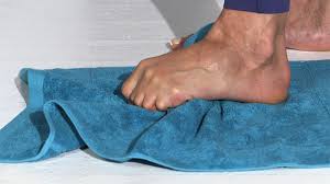
Towel Scrunches exercise

Towel Scrunches exercise


One exercise is to sit down and place a rolled up towel under your foot. While keeping your foot flexed, slowly roll the towel back and forth under your foot for a few minutes. This will help stretch and massage the plantar fascia in your foot.

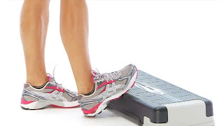
Calf Stretching Exercise


Calf Stretching Exercise

Another exercise you can do is to stand on a step and let your heel hang off the edge. Gently stretch your foot by moving your heel up and down. You should feel a stretch in your Achilles tendon. Hold this stretch for a few seconds and then release. Repeat a few times.


Ergonomics of Heel Pain:

Heel pain is a common issue that can be caused by a variety of factors. Poorly fitting shoes, overuse, and injury are all common causes of heel pain.

There are a few things you can do to help prevent heel pain. First, make sure you wear shoes that fit properly and support your feet. 

Secondly, avoid overdoing it when you are participating in activities that put stress on your feet and heels. If you do start to experience heel pain, be sure to rest and give your feet a break.

If you are suffering from heel pain, there are a few things you can do to find relief.  Pain relieving medication and oil can help to ease the pain. You can also try icing the affected area for 20 minutes at a time. If the pain is severe, you may need to see a doctor or physical therapist for more aggressive treatment.

Tuesday, 10 May 2022

Pull in Calf muscle: Cause, Symptoms, Treatment, Exercise

Pull in Calf muscle
Pull in Calf muscle

What is a pull in calf muscle?

A pull in calf muscle is also called calf muscle strain, it occurs when the muscles in the calf (the soleus and gastrocnemius) get overstretched during performing any activity of leg.

The calf muscles are located in the lower leg behind the shin bone and extend from the base of the thigh down to the heel. They work as flex and bend the foot, ankle, and knee. A calf strain can cause pain and make  difficulty to perform running, jumping, and doing other activities.

A pull in calf muscle can result in partial tear or complete tears depends upon level of injury. A torn calf muscle may require surgery to repair it.

What is the difference between muscle strain or muscle pull or a sprain?

Pull in Calf muscle also you can say A calf strain but it isn’t a sprain. Strains mean injuries to muscles or tendons (tissues that attach muscles to bones) while Sprains occurs injuries to ligaments (tissue that connects bones or cartilages or holds a joint along).

Where is the calf muscle located?

The calf muscle is a group of two main muscles — the gastrocnemius muscle, the soleus muscle and additionally plantaris muscle.

These two muscles the gastrocnemius and soleus muscles are collectively big muscles with two sections.

a small muscle called the plantaris travels between the gastrocnemius and soleus muscle throughout the length of the lower leg. these 3 muscles are also called the triceps surae. however, not everyone have a plantaris muscle. Mostly 100 % of individuals have 2 larger muscles.

Gastrocnemius Muscle is the larger Calf Muscle Forming The bulge of Muscle mass and visible beneath the skin. Gastrocnemius has 2 heads which looks like Diamond shape.

The Soleus Muscle is a Small Muscle, Flatt and located underneath The Gastrocnemius Muscle.

A majority of soleus muscle fibers originate from each side of the anterior aponeurosis, attached to the tibia and fibula.

Insertion:

Both Muscle Insertion merge via Tendo Achiilis and the Achilles tendon inserts into the heel bon(calcaneus).

Action:

Calf Muscle is one of strong lower limb Muscle which Planter Flex from Ankle Joint. 

This muscle is anti-gravity Muscle and normally all anti gravity Muscle require high Power that's Why All anti gravity Muscles are Bulky and Strong. 

Running, Jumping, Dancing, Cycling all this Activity uses Calf Muscle. 

Jumping is the activity where Highest uses Calf Muscle.

Who gets Pull in Calf muscle?

Anyone can have a pull in calf muscle. But these pull are most commonly seen in sports person who perform lots of stop-and-go movements with jerky bursts at ankle such as Sprinters, football players, Hockey players, soccer players, and tennis players are at risk of calf muscles strain. eventually, the injury is also called “tennis leg.”

What is the causes of pull in calf muscles?

Call muscle pull mostly occur when someone suddenly moves or overstretches in the calf muscle during moving or playing activity such as Fast pivots, jumps, or abrupt stops can cause muscle strains. This injury is mainly common when ankle get forced upward (toward the body) and the ankle stretches the calf muscles down too rapidly.

Following are the most common cause of calf muscle pull, these are:

  • Not warming up correctly before sports or physical activity
  • Calf Muscle tightness (deficient flexibility)
  • Calf muscle weakness (deficient conditioning)
  • Overexercise or Over physical activity (Overexertion and fatigue)

Acute calf muscle pull occurs during Slip body, jump, run, throw something, raise something heavy, lift something while clumsy position.

Chronic calf muscle pull occurs during Sports like row, playing Hockey, tennis, golf, or baseball, poor posture, holding back or neck in an ungainly position for long periods of time, like after work at a desk.

Overuse:

Repeated ankle movement can lead to microtrauma of calf muscle will cause overuse calf muscle pull.

Don’t stretch or warm up prior to exercise: Stretching prior to calf exercise step by step will increase how much stress you place on muscles.

Calf muscle tightness: If your calf muscles are tight and not flexible to high intensity activity in full range then your calf muscles (and the fibers in them) are tighter, which build them additional risk to strains.

Other risk factors for calf muscles pull are:

Age: People over 40 may be more likely to have muscle strains during physical activity.

Sex: Men are more likely to get calf muscle pull as compared to females.

Calf muscle weakness: It’s important to do warm up and stretch your calf prior to any physical activity and condition muscles before the start of a sports activity.

The Calf muscle pull is classified into three grades:

Grade 1: It is the type of muscle strain that occurs once a overstretch causes very few micro-tears among the muscle fibers. It could also be ready to resume an activity without pain or with gentle discomfort. Mostly post activity tightness and aching, Full recovery take within 1 weeks. average time to return to sport 10-12 days. sign-pain on unilateral calf raise or hop.

Grade 2: This strain include a partial tear of the muscle fibers. you may acquire to limit activity, but full recovery takes around five to eight weeks. average time to return sport after 20 to 30 days. sign-pain with active plantar flexion, pain and weakness with resisted plantar flexion, loss of flexion.

Grade 3: severe injury and instantly pain in the calf, and involves a complete tear or rupture of the affected muscle fibers. Full recovery can take 3 to 4 months and, in some instances, surgery may even be required. average time to come sport 6 months after surgery. sign-inability to contract calf muscle may have the palpable defect, Thomson’s test positive.

What are the symptoms of a pulled calf muscle?

  • Difficulty stretching the calf muscles or standing on toes or limping during walking.
  • Calf Muscle pain when flexing ankle or pointing toes.
  • Difficulty in knee movement or painful knee movement.
  • Snapping or popping sensation in calf muscles.
  • Sudden pain, spasm in the back of lower leg.
  • Swelling in calf muscle and also Ankle swelling.
  • Bruising on calf area.

Most people with a pulled calf muscle shows that not able to do their regular activity instantly following the injury.

Differential diagnosis:

  • Medial leg bone stress syndrome
  • Plantar fasciopathy
  • Ankle Joint sprain
  • Medial leg bone stress syndrome
  • Leg injuries related to sports with same symptoms and treatment as a calf strain are mentioned below.
  • Chronic exertional compartment syndrome (CECS)
  • TendoAchilles tendinopathy
  • Popliteal Artery entrapment Syndrome (PAES)

Diagnosis and tests:

You may require imaging study to checkout if there are any partial or complete tears in calf. These tests can also rule out other conditions that related lower leg pain, such as Achilles tendon ruptures or deep vein thrombosis (DVT).

Ultrasound, to examine for calf tears or fluid buildup within the calf muscles.
MRI, to examine for blood clots, a tear, or internal bleeding.

Assessment of lower leg:

Subjective assessment

Ask the patient pain history with associated symptoms
mechanism of injury:
Inciting trauma- side and magnitude of injury force
recurrent trauma-faulty postural related injuries

Objective assessment

Examine the foot and ankle in supine and standing position
Ankle Active range of motion
Ankle Passive range of motion
Resisted strength check the ankle and foot
Knee Active ROM and manual muscle testing

On objective assessment, there will be:

muscle Tenderness- to touch at the point of injury
Swelling
Bruising may appear within hours or days
Stretching of the muscle will reproduce pain
Pain on resisted plantar flexion.

Treatment of Pull in Calf muscle:

Medical Treatment

Calf muscle pull treatment mostly depends upon grade of injury. Grade 1 or Grade 2 muscle strain recover with few days rest with Pain relieving medicine mostly Non-Steroidal Anti-Inflammatory Drugs (NSAIDs) and use of RICE Principle.

RICE Principle are:

R- rest
I- ice for cooling
C- compression: tapping and splinting
E- elevation

Few days rest helps to recover calf muscle strain to heal properly while ice pack helps to relieve pain and swelling.

Elevation of limb is required if Ankle swelling is present otherwise not required.
Crep bandage or taping also helps to stabilize calf muscles.

After few days rest pain will disappear and other related symptoms such as muscle spasm, swelling is relived exercise is the best option to return to normal activity.

Physiotherapy Treatment:

Physiotherapist will assess your condition and accordingly start the treatment depends upon severity of injury and symptoms.

Mostly Physiotherapist uses pain relieving Electrotherapy modalities such as Transcutaneous electrical nerve stimulation (TENS), Interferential therapy(IFT), Ultrasound therapy machine. This electro. modalities helps to relieve pain, swelling and muscle spasm.

Physiotherapist teaches you initially light exercise and as pain and other symptoms relive they give you stretching and strengthening exercise of calf muscles.

Pull in calf muscle rehabilitation exercises:

Phase one- the 1st week after injury

Use of ice pack 3 times par day
Rest
Avoid exercise or do only isometric pain free exercise.

Phase two- the 2nd and 3rd weeks after injury

Start active Ankle range of motion (ROM) exercises
Avoid painful activities.
range of motion exercise: knee flexion exercise
knee extension
plantar flexion-dorsiflexion
genital pain free calf resisted exercise
Gradual pain free calf stretching exercises
Use of ice appliance 15-20 min, after exercise

Phase three-3 to 4 weeks after injury

Gentle strengthening exercises:
knee flexion-extension
plantar flexion- dorsiflexion
gental pain-free calf stretching
you can also start with thera-band resisted calf exercise(pain free)

Following are the few best exercise you can start at home in pain free manner.

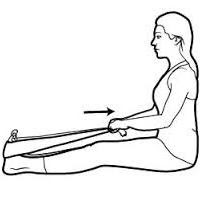
1. Towel calf stretch:
Towel calf stretch
Towel calf stretch


Sit on a firm mat with one leg straight ahead of you. Loop towel circling your ankle as per picture and pull the towel toward body keeping knee straight. now hold this position for 15 to 30 seconds and then relax. Repeat 3 times.

2.Seated heel Raises:

Take a sitting position on a chair with both legs on the ground. Pressing down through the toes, raise heels off the floor.

Hold this position for 5 to 10 seconds.Repeat 10-15 times.

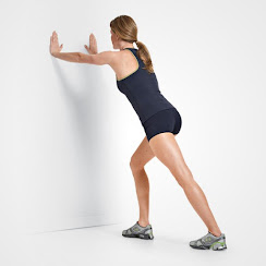
3.Standing calf stretch:
Standing calf stretch

Keep your both leg in same line position with around minimum foot distance with forward leg flexed from Knee and back leg straight , You will feel stretching in back of lower leg in Calf muscle and Hold for few seconds then relax. exercise must be pain free and gentle stretch initially for few days.

Monday, 4 April 2022

Ankle pain without Injury: Cause, Symptoms, Treatment, Exercise

Ankle pain without Injury

If you have Sudden Ankle pain without injury, there may too many cause of this pain and symptoms are vary according to conditions. Treatment of Ankle pain is also depends upon cause, symptoms and diagnosis of Ankle pain.

In this article we discuss what are the possible cause of Ankle pain without Injury and symptoms. we also discuss how to diagnose ankle pain, Medical treatment and Physiotherapy exercise.

What are the causes of Ankle pain without injury?

There are too many causes of Ankle pain still if you are not injured, few most common cause of Ankle pain are:

  • Overuse of Ankle joint- continuous work load mainly farmers and laborers require 8 to 10 hours work in standing and walking position, these may lead to pressure on the joint and causes pain
  • osteoarthritis of Ankle joint: These are other cause of Ankle pain in which age related wear and tear of the Ankle joint may leads to damaged to joint surface.
  • Bursitis: Bursae (Fluid-filled sacs) arount the joint which provides cushion to your bones when they move. When bursae become irritated and inflamed called Bursitis.
  • Tendonitis: Muscle's tendon become irritated, inflamed called tendonitis. These tendons connect muscles to bones. Continuous workload may lead stress on the muscle's tendon such as Achilles tendinitis.
  • Plantar fasciitis causes pain in the bottom of the heel. The plantar fascia is a thin ligament that connects your heel to the front of your foot. It supports the arch in your foot and is important in helping you walk.
  • Flatfoot: When your arch is not properly develop from birth called congenital flatfoot which may lead to pain and swelling in the ankles and feet.
  • Gout: When uric acid increases in your body, causes multiple joint pain including Ankle joint. These uric acid created crystals which are deposited in the joint and causes joint pain, swelling and also restrict the movement most affect both the Ankles.

What are the signs and symptoms of ankle pain without injury?

  • Ankle pain with swelling
  • Bruising
  • Redness
  • Numbness or tingling
  • Instability
  • Ankle Joint Pain
  • Inability to bear weight on the affected ankle
  • Ankle joint Stiffness
  • Weakness
  • Discoloration
  • Warmth
  • Tenderness
  • Difficulty in walking
Diagnosis Ankle pain without Injury:

During a physical examination, the Physiotherapist will examine the patient’s ankle, foot, and lower limb. The doctor will also check the area of skin around the injury for proper points of tenderness and move the patient’s Ankle to check the range of motion and to know what positions cause discomfort or pain. 

Following are the imaging study also helpful for differential diagnosis of Ankle pain:

X-ray: X-Ray is mainly helpful if any arthritis changes are seen in the joint.
Magnetic resonance imaging (MRI): If x-ray is not helpful then further investigation Doctor recommend you MRI which uses radio waves & a strong magnetic field to produce detailed cross-sectional or 3-D images of the soft internal tissue of the ankle, with ligaments.
CT scan: If Doctor can get more detail view regarding the bones of the joint. CT scan takes too X-rays from many variant angles and mixes them to make cross-sectional or 3-D images.
Ultrasound: An ultrasound uses sound waves to produce images. These images may help doctors find the condition of a ligament/tendon when the foot is in a different position.

Treatment of Ankle pain without injury:

Medical treatment:

Mostly your Doctor prescribes you pain relieving medicine mostly NSAIDs are most common such as Diclofenac, Aceclefenac are enough to manage the ankle pain with Pain relieving gel and patches.

Home remedies for Ankle pain you can do is mainly use of Hot Packs- Cold Packs that helps to relieve pain and swelling naturally.

Physiotherapy treatment:

Physiotherapy treatment are mostly depends upon cause, symptoms and diagnosis of your Ankle pain. Most commoly Physiotherapy treatment in Ankle pain are pain relieving Electrotherapy modalities and Exercise.

Electrotherapy treatment:

There are various electro. modalities are uses to relive pain, swelling mainly in ankle pain three modalities would be beneficial which are describe below:

Ultrasound: Ultrasound is usually apply on tender point to relieve of pain.
TENS (Transcutaneous electrical nerve stimulation): TENS modality is mainly for reduction of pain, swelling.
IFT (Interferential Therapy ): If patient have any type of swelling, pain, inflammation ,IFT would be most effective modality.

Exercises for Ankle pain:

Regular exercise must help the patient to relieve pain and strengthen weak muscles.

Stretching exercise:

Achilles tendon & plantar fascia stretch:

To do this exercise, you can loop a towel around the ball of your foot & pull it back towards your body with a straight knee. Hold for 10 to 20 seconds for each foot and do it 3 times.

Sitting plantar fascia stretch:

To do this exercise you should cross your foot over the opposite knee. The base of the toes is put on the ground & gently pull back until you feel a stretch along the innerside of your foot. Hold for 10 to 30 seconds per repetition. Repeat 3 times on each foot.

Plantar fascia stretch:

To do this exercise you should put a towel on the floor in front of your chair, with the heels which are on the ground, try to pick up the towel by grabbing it with your toes, this exercise Repeat 10-15 times with 2 times a day.

Standing calf stretch:
Standing calf stretch

To do this exercise, you must stand facing a wall then place both hands on the wall which is in front of you. Put one foot forward to the other with the front foot about 30cm away from the wall, keep the other knee straight with keep your back heel flat on the floor. Gradually bend the front knee until you feel gentle stretch in the calf of the back lower leg. Relax and repeat 8 to 10 times. This exercise may also be done with the back knee slightly bent for a milder stretch.

Deep calf stretch:

To do this exercise you must stand on a step with the heel hanging off the edge. Your may also allow holding onto something for support. You should gradually drop heel towards the floor and feel a stretch in the calf muscles & Achilles tendon. Hold this for 10 to 30 seconds. Repeat this exercise 2-3 times.