Showing posts with label Shoulder Pain. Show all posts

Monday, 11 March 2024

Trapezius Muscle Pain

What is a Trapezius Muscle Pain?

Trapezius muscle pain

Trapezius muscle pain can range from mild discomfort to severe, debilitating pain, affecting daily activities and overall quality of life.

One of the main muscles of the back and neck is the trapezius muscle, so named because of its trapezoidal shape. It reaches across the shoulder blades and descends from the base of the skull to the thoracic spine. This large muscle is important for many movements of the upper back, shoulders, and neck, such as pulling, lifting, and rotating the shoulders.

Despite the trapezius muscle's strength and durability, it can become painful or uncomfortable for a variety of reasons, including bad posture, overuse, stress, or injury.

It is essential to understand the causes, signs, and effective treatments of trapezius muscle pain in order to manage this common condition while improving musculoskeletal health. This article discusses the trapezius muscle's anatomy and function, common causes of pain in the area, warning signs, and preventative and therapeutic measures.

Related Anatomy

Trapezius Muscle Anatomy

The trapezius muscle is a large, superficial muscle that covers much of the upper back and neck. It is divided into three distinct regions: the upper (or superior), middle, and lower (or inferior) trapezius.

Upper Trapezius:

Origin: Occipital bone (base of the skull) and the spinous processes of the cervical vertebrae (C1-C4).

Insertion: Clavicle (collarbone) and acromion process of the scapula (shoulder blade).

Function: Elevates and upwardly rotates the scapula. It also assists in neck extension and lateral flexion.

Middle Trapezius:

Origin: Spinous processes of the thoracic vertebrae (T1-T5).

Insertion: Medial aspect of the acromion process and superior lip of the spine of the scapula.

Function: Retracts (adducts) the scapula, pulling it toward the spine.

Lower Trapezius:

Origin: Spinous processes of the thoracic vertebrae (T6-T12).

Insertion: Medial end of the spine of the scapula.

Function: Depresses (pulls downward) and upwardly rotates the scapula. It also assists in scapular retraction.

Nerve Supply:

The trapezius muscle is innervated by the spinal accessory nerve (cranial nerve XI) and branches of the cervical spinal nerves (C3-C4).

Blood Supply:

Blood is supplied to the trapezius muscle primarily by branches of the superficial cervical artery, transverse cervical artery, and dorsal scapular artery.

To understand the function of the trapezius muscle and the possible causes of pain and dysfunction, one must have a thorough understanding of its complex anatomy. Any area of the trapezius muscle that is dysfunctional can cause pain, restricted movement, and poor posture. For this reason, it is critical to properly assess the situation and implement targeted interventions to address problems related to the trapezius muscle.

Trapezius Muscle Pain Causes

Causes of Trapezius Pain are:

Poor Posture:

Extended periods of sitting or standing with bad posture can cause the trapezius muscle to become overworked and strained. Muscle soreness and discomfort can arise from the trapezius becoming overstretched and fatigued from slouching or hunching the shoulders forward.

Muscle Overuse

Overuse of the trapezius muscle can result from repetitive motions or activities involving the arms, shoulders, and neck. This usually happens in jobs or activities where lifting, carrying, or reaching overhead for extended periods of time is required.

Muscle Tension and Stress

People may unintentionally tense their necks and shoulders in response to emotional stress, anxiety, or tension, which can increase the tension in the trapezius muscle. Trigger points and muscle soreness can arise as a result of persistent muscle tension.

Trauma or Injury

Acute pain and inflammation can be brought on by direct trauma to the trapezius muscle, such as a fall or impact, which can result in muscle strains, tears, or contusions. Furthermore, over time, repetitive microtrauma from manual labor or sports-related activities can result in chronic muscle injuries.

Neck and Shoulder Strain

Strains to the muscles, ligaments, or joints of the neck and shoulders can cause pain that refers to the trapezius muscle. Secondary trapezius muscle pain can be caused by diseases like cervical spondylosis, whiplash injuries, or shoulder impingement syndrome.

Poor Ergonomics

Pain in the trapezius muscle can be caused by a poorly designed piece of equipment, incorrect lifting techniques, or an incorrectly set up workstation. It's crucial to maintain good ergonomics to avoid tense and sore muscles when performing daily tasks.

Nerve Compression

Referred pain, tingling, or numbness in the muscle can be caused by compression or irritation of the spinal accessory nerve or cervical spinal nerves, which supply the trapezius muscle.

Postural Imbalances

Unbalances in the muscles that surround the trapezius, like those in the pectoral or rhomboids, can cause compensatory movements and put more strain on the trapezius, making it more vulnerable to pain and dysfunction.

Symptoms of Trapezius Muscle Pain

Other Associated Symptoms are:

Shoulder and Neck Discomfort

Trapezius muscle pain frequently manifests as neck, shoulder, and upper back pain. The pain can be dull, achy, or sharp, and its severity ranges from mild to severe.

Muscle Tenderness

When the trapezius muscle is palpated, certain trigger points or the muscle fibers themselves may be tender or tense in certain places.

Restricted Range of Motion

Pain in the trapezius muscles can restrict shoulder and cervical range of motion, making it challenging to execute actions like raising arms overhead, turning the head, or twisting the neck.

Muscle Stiffness and Tightness

Muscle stiffness and tightness are common symptoms of trapezius muscle pain, especially in the upper back and neck. Feelings of discomfort and decreased flexibility may result from this.

Headaches

Tension headaches or cervicogenic headaches can result from pain that radiates from the trapezius muscle to the head and temples. The dull, band-like sensation that surrounds the head is a common symptom of these headaches.

Shoulder Blade Pain

Pain may be localized to the area between the shoulder blades (scapulae), particularly when the middle and lower trapezius muscles are involved.

Trapezius Muscle Spasms

In rare cases, involuntary muscle contractions or spasms may accompany pain in the trapezius muscle, making movement more difficult and uncomfortable.

Numbness or Tingling

Numbness, tingling, or pins and needles may be experienced in the affected area due to compression or irritation of the nerves innervating the trapezius muscle.

Postural Changes

 People who experience chronic pain in the trapezius muscle may adopt compensatory postures or movements to ease their discomfort. As a result, there may be more musculoskeletal imbalances and pain throughout time.

Fatigue and Weakness

It may be difficult to carry out daily tasks involving the shoulders and neck when dealing with chronic pain in the trapezius muscle.

Differential Diagnosis

When making a differential diagnosis for pain in the trapezius muscle, other medical conditions that may have similar symptoms are taken into account. Among the possible differential diagnoses are the following:

  • Cervical Radiculopathy: Pain, numbness, or tingling that radiates from the cervical spine and may resemble pain in the trapezius muscle is caused by compression or irritation of the spinal nerves. Cervical radiculopathy can cause weakness or changes in sensation in the upper extremities, and it is frequently accompanied by neck pain.
  • Rotator Cuff Injury: Damage to the rotator cuff's muscles and tendons in the shoulder can result in pain and a restricted range of motion that can be felt in the neck and upper back. People who have experienced shoulder trauma or who perform repetitive overhead activities are more likely to sustain rotator cuff injuries.
  • Cervical Disc Herniation: Herniation of the intervertebral discs in the cervical spine can compress surrounding nerves, causing pain in the neck, shoulders, and arms. Herniated cervical discs can also result in abnormal reflexes, sensory abnormalities, and muscle weakness.
  • Thoracic Outlet Syndrome (TOS): The condition known as Thoracic Outlet Syndrome (TOS) is caused by compression of nerves or blood vessels in the thoracic outlet, which is the area between the collarbone and the first rib. TOS symptoms include arm and shoulder pain, tingling, numbness, and weakness. Certain activities or postures that compress the thoracic outlet can make TOS worse.
  • Fibromyalgia: This is a chronic pain syndrome that is marked by body-wide musculoskeletal pain, fatigue, and tender points. Although fibromyalgia can include pain in the trapezius muscle, the illness usually presents with several tender points and systemic symptoms.
  • Myofascial Pain Syndrome: Referred pain patterns can be caused by the presence of trigger points, which are small, tender, spastic areas of muscle. Although the symptoms of trigger points in the trapezius muscle can resemble those of trapezius muscle pain, they can be distinguished by manual palpation and trigger point identification.
  • Postural Syndromes: Musculoskeletal pain in the upper back, neck, and shoulders can result from long-term poor posture or anatomical abnormalities in the spine or shoulders. Managing postural-related pain requires addressing underlying postural imbalances and ergonomic issues.
  • Shoulder Impingement Syndrome: Shoulder pain and dysfunction that may be referred to the trapezius muscle area can be caused by impingement of the rotator cuff tendons or bursa between the humeral head and the acromion process of the scapula.

Diagnosis

A thorough evaluation by a medical professional is usually necessary to diagnose trapezius muscle pain. This evaluation may consist of the following elements:

Medical History: The healthcare professional will start by getting details regarding the patient's symptoms, such as where, when, and how much pain they are experiencing, along with any triggers or mitigating factors. A thorough medical history aids in determining any possible contributing factors or underlying causes.

Physical Examination: To evaluate the flexibility, strength, and range of motion of the shoulders, neck, and upper back, a comprehensive physical examination is performed. To find trigger points, tight spots, or tender spots in the trapezius muscle, the medical professional may palpate the muscle.

Neurological Examination: To assess nerve function, reflexes, and sensory function in the upper limbs, a neurological examination may be carried out. This aids in the evaluation of any indications of nerve irritation or compression that might be causing the symptoms.

Imaging Studies: X-rays, CT (Computed Tomography) scans, and MRIs (Magnetic Resonance Imaging) may occasionally be requested in order to rule out underlying structural abnormalities or injuries in the neck, shoulders, or spine. These imaging modalities can assist in locating any possible sources of pain or dysfunction as well as provide detailed images of the musculoskeletal structures.

Nerve Conduction Studies and Electromyography (EMG): These tests may be suggested to evaluate the electrical activity and function of the muscles and nerves that supply the trapezius muscle. Neurological disorders and abnormalities of the muscles can be diagnosed with the aid of nerve conduction studies and EMG.

A diagnosis of trapezius muscle pain or another musculoskeletal condition may be made in according to the evaluation's results. The recommended course of treatment will be individualized to target the particular underlying cause and reduce symptoms; this may include a mix of complementary therapies, medication, physical therapy, and lifestyle changes.

Treatment of Trapezius Muscle Pain

A combination of conservative treatments targeted at pain reduction, muscle function enhancement, and addressing underlying contributing factors is usually used to treat trapezius muscle pain. 

Common treatment options are:

Rest and Activity Modification: Reducing activities that aggravate pain in the trapezius muscle or taking a brief break from them can help manage symptoms and stop the muscle from getting worse. It's crucial to stay out of positions or repetitive movements that put strain on the trapezius muscle during the first stages of treatment.

Physical therapy: A customized program for physical therapy can help improve flexibility, strengthen the trapezius muscle, and address postural imbalances. To lessen discomfort and accelerate healing, practitioners can use manual therapy, stretching exercises, therapeutic exercises, and modalities like TENS, IFC Machine, heat or ice therapy.

Pain management: Nonsteroidal anti-inflammatory medications (NSAIDs) available over-the-counter, such as ibuprofen or naproxen, can help lessen pain and inflammation caused by pain in the trapezius muscle. For temporary pain relief, doctors may occasionally prescribe prescription drugs or muscle relaxants.

Trigger Point Therapy: Tightness and tension in the trapezius muscle can be released with the use of manual techniques like trigger point massage, myofascial release, or dry needling. Localized pain relief and improved muscle function can be achieved by focusing on trigger points located within the muscle.

Posture Correction: Preventing recurrent pain in the trapezius muscle requires addressing underlying postural imbalances and ergonomic factors. It might be advised to provide education on good posture for everyday tasks, workplace ergonomics, and ergonomic adjustments.

Stress management: By promoting relaxation and easing muscle tension, stress-reduction methods like yoga, deep breathing exercises, mindfulness meditation, and relaxation exercises can lessen the chance of aggravating trapezius muscle pain.

Therapeutic modalities can be used to relieve pain, lower inflammation, and encourage tissue healing in the trapezius muscle. These include heat therapy, cold therapy, ultrasound, and electrical stimulation.

Lifestyle Changes: Adopting healthful routines like consistent exercise, enough sleep, a balanced diet, and adequate hydration can promote general musculoskeletal health and lower the incidence of pain and dysfunction in the muscles.

Ergonomic Modifications: Changing the way that workstations, chairs, computers are set up, and lifting is done can help lessen the strain that regular activities place on the trapezius muscle and lower the chance of developing muscle soreness.

Injections: For localized pain relief and to reduce inflammation in the trapezius muscle, corticosteroid injections or trigger point injections may be considered in certain situations.

Physiotherapy Treatment of Trapezius Muscle Pain

Physiotherapy is essential for treating pain in the trapezius muscle because it corrects muscle imbalances, restores mobility, increases strength, and encourages good posture. 

The following elements of a physiotherapy treatment plan for pain in the trapezius muscle may be available:

Manual Therapy Techniques:

  • Soft tissue mobilization: A massage technique used to ease the trapezius muscle's tightness and tension.
  • Joint mobilization: Mild manual techniques to ease shoulder and neck stiffness and increase joint mobility.

Therapeutic Exercises:

  • Stretching exercises: Focused stretches to increase flexibility and decrease muscle tension in the trapezius muscle.
  • Strengthening exercises: Postural muscles are the emphasis of these progressive resistance workouts, which aim to strengthen the trapezius and surrounding muscles and support good alignment and stability.
  • Scapular stabilization exercises: These exercises help to prevent shoulder impingement and lessen strain on the trapezius muscle by enhancing scapular control and muscle balance.

Postural Correction:

Instruction on ergonomic adjustments, daily activities, and work tasks that promote good posture to avoid straining the trapezius muscle.

exercises for postural retraining to enhance awareness and alignment of the head, shoulders, and spine.

Modalities:

Applying heat packs or warm compresses to the trapezius muscle to improve blood flow, ease tension, and reduce pain is known as heat therapy.

Cold therapy: Applying ice or cold packs to the trapezius muscle to relieve pain and reduce inflammation.

Electrical stimulation: To reduce pain and encourage muscle relaxation, neuromuscular electrical stimulation (NMES) or transcutaneous electrical nerve stimulation (TENS) may be utilized.

Trigger Point Treatment:

By deactivating trigger points in the trapezius muscle, trigger point release techniques—such as manual pressure, ischemic compression, or dry needling—can lessen referred pain patterns.

Learning and Self-Control:

Patient education regarding ergonomics, good body mechanics, and ways to avoid straining muscles and having trapezius muscle pain recurrence.

guidance on self-care practices, stretching regimens, and at-home workouts to speed up recovery and preserve musculoskeletal health.

Gradual Rehabilitative Intervention:

Progressive increase in activities and exercises as function improves and pain subsides, emphasizing everyday living activities and functional movements.

Monitoring development and modifying the treatment plan in accordance with each patient's needs and preferences.

An comprehensive physiotherapy strategy catered to the requirements and objectives of the patient can successfully treat pain in the trapezius muscle, restore function, and enhance quality of life. To maximize results and achieve long-term musculoskeletal health, the patient and physiotherapist must collaborate and communicate on a regular basis.

Exercises for Trapezius Muscle Pain

The goals of exercises for pain in the trapezius muscle are to reduce stress, enhance muscle strength and flexibility, and encourage good posture.
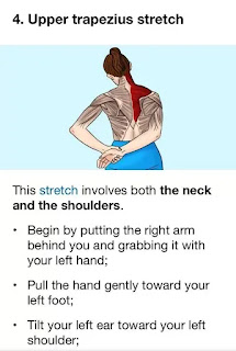
Upper Trapezius Stretch

The following efficient exercises can help reduce pain in the trapezius muscle:

Upper Trapezius Stretch:

  • Keep your back straight and sit or stand tall.
  • Till you feel a stretch along the side of your neck, gently tilt your head to one side and bring your ear towards your shoulder.
  • After holding the stretch for 15 to 30 seconds, move on to the opposite side.
  • Two to three repetitions per side.

Levator Scapulae Stretch:

  • Take a tall stance or sit upright, then slowly turn your head to the side and look over your shoulder.
  • Place your hand on the top of your head and apply gentle pressure to increase the stretch.
  • Hold the stretch for 15-30 seconds, then switch to the other side.
  • Repeat 2-3 times on each side.

Scapular Retraction:

  • Stand with your back against a wall or in a neutral position.
  • As though you were attempting to hold a pencil between your shoulder blades, squeeze them together.
  • Hold the squeeze for 5-10 seconds, then relax.
  • Repeat for 10-15 repetitions.

Shoulder Shrugs:

  • Stand or sit with your arms at your sides.
  • Shrug your shoulders upwards towards your ears as high as possible.
  • Hold for 1-2 seconds, then lower your shoulders back down.
  • Repeat for 10-15 repetitions.

Trapezius Strengthening with Resistance Bands:

  • A stable object at waist height should be used to fasten one end of a resistance band.
  • Hold the other end of the band in one hand and stand with your side facing the anchor point.
  • Keep your arm straight and pull the band diagonally across your body, engaging the trapezius muscle.
  • Slowly return to the starting position.
  • Perform 10-15 repetitions on each side.

Neck Retraction Exercise:

  • Maintain a neutral head posture while sitting or standing.
  • Gently tuck your chin towards your neck, creating a double chin.
  • Hold for a few seconds, then release.
  • Repeat for 10-15 repetitions.

Wall Angels:

  • Stand with your back against a wall and your arms bent at 90 degrees with your elbows and wrists touching the wall.
  • Slowly slide your arms upwards along the wall while keeping your elbows and wrists in contact with the wall.
  • Squeeze your shoulder blades together the entire time you perform the movement.
  • Return your arms to the starting position by sliding them down.
  • Repeat for 10-15 repetitions.

Thoracic Extension Stretch:

  • Sit on the floor with a foam roller positioned horizontally under your upper back.
  • Support your head with your hands, keeping your elbows bent.
  • Slowly lean back over the foam roller, arching your upper back and extending your thoracic spine.
  • After holding the stretch for 15 to 30 seconds, go back to your starting posture.
  • Repeat 2-3 times.

Regularly perform these exercises, paying attention to your form and deliberate movements. Stretch lightly at first, then progressively increase the resistance and intensity as tolerated. Before beginning any new exercise program, especially if you have pre-existing health conditions or concerns, get advice from a medical professional or physical therapist.

Keeping a healthy lifestyle, practicing proper posture, and putting ergonomic techniques into practice can all help prevent pain in the trapezius muscle. 

The following advice can help avoid pain in the trapezius muscle:

Maintain Proper Posture:

Maintain a straight back, relaxed shoulders, and a head that is in line with your spine when you sit or stand.

Stay away from hunching forward or slouching as these postures can strain the trapezius muscle and cause imbalances in the muscles.

Take Regular Breaks:

Take frequent breaks to stand up, stretch, and move around if your job requires you to sit at a desk for extended periods of time or if it's sedentary.

Throughout the day, gently stretch your shoulders and neck to release tension and avoid stiffness.

Practice Ergonomic Work Habits:

To encourage neutral posture and reduce strain on the trapezius muscle, rearrange your workspace.

In order to lessen neck strain, place your computer monitor at eye level and use an ergonomic chair with adequate lumbar support.

Benefit from ergonomic add-ons like supportive cushions, adjustable desks, and ergonomic keyboards.

Use Proper Lifting Techniques:

Use your leg muscles to lift heavy objects instead of your back and shoulders by bending your knees and maintaining a straight back.

When lifting, keep the object close to your body and try not to twist or reach awkwardly.

Stay Active and Exercise Regularly:

Exercise on a regular basis to build strength in the muscles of your neck, shoulders, and upper back, especially the trapezius muscle.

Include activities that help you maintain good posture, like core and back strengthening exercises.

Practice exercises that promote flexibility and mobility, like yoga, Pilates, or swimming.

Manage Stress and Tension:

To release tension in your shoulders and neck, try stress-reduction methods like deep breathing, meditation, or mindfulness.

Throughout the day, take breaks to unwind and reduce stress, particularly if you're dealing with a heavy workload or emotional strain.

Drink plenty of water and eat a balanced diet:

In order to keep muscles hydrated and avoid dehydration, which can exacerbate tension and cramping in the muscles, drink lots of water throughout the day.

To enhance overall muscle health and function, maintain a well-balanced diet high in vitamins, minerals, and nutrients.

Get Adequate Sleep:

Make sure you are getting enough restorative sleep every night by making quality sleep a priority.

Invest in pillows and a supportive mattress to help maintain healthy spinal alignment and lessen shoulder and neck strain when you sleep.

These preventive steps can help lower your chance of experiencing pain in your trapezius muscles and improve your overall musculoskeletal health.

Summary

In summary, pain in the trapezius muscle is a common condition that can greatly affect everyday activities and quality of life. Effective management and prevention of trapezius muscle pain require an understanding of its anatomy, causes, symptoms, and available treatments. The most common causes of pain in the trapezius muscle are injury, stress, poor posture, and overuse of the muscles.

Physiotherapy is essential for reducing pain in the trapezius muscle and enhancing muscle function and mobility. It includes manual therapy techniques, therapeutic exercises, and modalities. Furthermore, preventing trapezius muscle pain can be achieved by putting preventive measures into practice, such as maintaining good posture, adopting ergonomic work habits, being active, controlling stress, and getting enough sleep.

Through the implementation of focused interventions and a comprehensive approach to musculoskeletal health, people can effectively manage pain in the trapezius muscle, enhance their general quality of life, and avoid further episodes of pain. For a customized assessment and treatment recommendations, it's crucial to consult a healthcare provider if you suffer from severe or chronic muscle pain. People may achieve optimal musculoskeletal health and long-term relief through preventive care and lifestyle adjustments.

Tuesday, 28 March 2023

Shoulder Impingement Syndrome: Cause, Symptoms, Treatment, Exercise

What is a Shoulder impingement syndrome?

Shoulder Impingement Syndrome

Shoulder impingement syndrome (SIS) is a condition where the soft tissues in the shoulder joint become compressed or pinched, causing pain, inflammation, and limited movement. This compression typically occurs in the subacromial space, which is the narrow passageway between the acromion (a bony protrusion at the top of the shoulder blade) and the rotator cuff tendons.

The rotator cuff is a group of four muscles and their tendons that surround the shoulder joint and help to stabilize and move the shoulder. When the rotator cuff tendons become inflamed or irritated, they can swell and thicken, making it more difficult for them to move smoothly through the subacromial space. This can lead to rubbing and compression of the tendons and other soft tissues, causing pain and discomfort.

SIS can be caused by a variety of factors, including overuse, poor posture, muscle imbalances, and structural abnormalities in the shoulder joint. It is a common condition among athletes, particularly those who engage in overhead activities such as throwing, swimming, or weightlifting.

Treatment for SIS typically involves rest, physical therapy, and anti-inflammatory medications. In more severe cases, surgery may be necessary to relieve compression in the subacromial space.

Related Anatomy

To understand shoulder impingement syndrome (SIS), it is important to have a basic understanding of the anatomy of the shoulder joint.

The shoulder joint is a ball-and-socket joint that connects the upper arm bone (humerus) to the shoulder blade (scapula). The ball of the joint is formed by the rounded end of the humerus, while the socket is formed by a shallow depression on the scapula called the glenoid fossa. The joint is stabilized by a group of muscles and tendons called the rotator cuff, which wraps around the head of the humerus and helps to hold it in place within the socket.

The subacromial space is a narrow passageway between the acromion, a bony projection of the scapula, and the rotator cuff tendons. The space is lined with a lubricating sac called the subacromial bursa, which helps to reduce friction as the tendons move through the space. When the rotator cuff tendons become inflamed or irritated, they can swell and thicken, making it more difficult for them to move smoothly through the subacromial space. This can lead to rubbing and compression of the tendons and other soft tissues, causing pain and discomfort.

Other important structures in the shoulder joint include the labrum, a ring of cartilage that helps to deepen the socket and provide additional stability to the joint, and the biceps tendon, which attaches the biceps muscle to the shoulder blade and helps to stabilize the joint during certain movements.

What are the causes of Shoulder impingement syndrome?

Shoulder impingement syndrome (SIS) can be caused by a variety of factors, including:

  • Overuse: Activities that involve repetitive overhead movements, such as throwing a ball or painting a ceiling, can put a lot of stress on the rotator cuff tendons and lead to inflammation and irritation.
  • Poor posture: Poor posture can cause the shoulder joint to become misaligned, which can put extra pressure on the rotator cuff tendons and lead to impingement.
  • Muscle imbalances: Imbalances in the muscles that surround the shoulder joint can cause the joint to become unstable and increase the risk of impingement.
  • Structural abnormalities: Some people are born with structural abnormalities in the shoulder joint, such as a hooked acromion or a narrow subacromial space, which can increase the risk of impingement.
  • Trauma: A fall or other injury that causes damage to the rotator cuff tendons or other soft tissues in the shoulder can lead to impingement.
  • Aging: As we age, the tendons in the rotator cuff can become weaker and more prone to injury and inflammation, which can increase the risk of impingement.

Other conditions: Certain medical conditions, such as arthritis, can cause inflammation and swelling in the shoulder joint, which can lead to impingement.

Symptoms of Shoulder impingement syndrome

The symptoms of shoulder impingement syndrome (SIS) can vary depending on the severity of the condition, but common symptoms include:

  • Pain: Pain in the shoulder joint is the most common symptom of SIS. The pain is usually felt in the front or side of the shoulder and may be sharp or dull. The pain may be worse when the arm is lifted overhead or when the arm is moved away from the body.
  • Weakness: Weakness in the affected arm is another common symptom of SIS. This can make it difficult to perform certain activities that require lifting or reaching, such as putting on a shirt or reaching for a high shelf.
  • Limited range of motion: Impingement can limit the range of motion in the shoulder joint, making it difficult to move the arm in certain directions.
  • Clicking or popping sounds: Some people with SIS may experience clicking or popping sounds in the shoulder joint when they move their arm.
  • Swelling or inflammation: Inflammation and swelling in the shoulder joint may be present in more severe cases of SIS.
  • Pain at night: Some people with SIS may experience pain that wakes them up at night, particularly if they sleep on the affected side.

If you are experiencing any of these symptoms, it is important to see a healthcare professional for an accurate diagnosis and appropriate treatment.

Risk factor

There are several risk factors that can increase the likelihood of developing shoulder impingement syndrome (SIS), including:

  • Age: SIS is more common in older adults, as the tendons in the rotator cuff can weaken and become more prone to injury and inflammation with age.
  • Overuse: Repetitive overhead activities, such as throwing a ball or painting a ceiling, can increase the risk of SIS by putting a lot of stress on the rotator cuff tendons.
  • Poor posture: Poor posture can cause the shoulder joint to become misaligned, which can increase the risk of impingement.
  • Muscle imbalances: Imbalances in the muscles that surround the shoulder joint can cause the joint to become unstable and increase the risk of impingement.
  • Structural abnormalities: Some people are born with structural abnormalities in the shoulder joint, such as a hooked acromion or a narrow subacromial space, which can increase the risk of impingement.
  • Previous shoulder injuries: Previous injuries to the shoulder, such as a dislocated shoulder or a rotator cuff tear, can increase the risk of developing SIS.
  • Certain sports or activities: Athletes who engage in sports that require a lot of overhead movements, such as baseball or swimming, are at an increased risk of developing SIS.
  • Occupation: Jobs that require repetitive overhead activities, such as construction or painting, can increase the risk of developing SIS.
  • Genetics: Some people may be more predisposed to developing SIS due to genetic factors that affect the structure and function of the shoulder joint.

It's important to note that having one or more of these risk factors doesn't necessarily mean that someone will develop SIS, but it does increase their likelihood of developing the condition.

Differential Diagnosis

Shoulder impingement syndrome (SIS) can have similar symptoms to other conditions affecting the shoulder, so it's important to consider differential diagnoses when evaluating a patient with shoulder pain. Some potential differential diagnoses for SIS include:

  • Rotator cuff tear: A rotator cuff tear is a tear in one or more of the rotator cuff tendons that can cause pain, weakness, and limited range of motion in the shoulder joint.
  • Bursitis: Bursitis is inflammation of the bursae, small fluid-filled sacs that cushion the joints. In the shoulder, bursitis can cause pain, swelling, and limited range of motion.
  • Frozen shoulder: Frozen shoulder, also known as adhesive capsulitis, is a condition in which the shoulder joint becomes stiff and painful, often as a result of injury or surgery.
  • Shoulder arthritis: Arthritis in the shoulder joint can cause pain, stiffness, and limited range of motion in the shoulder.
  • Labral tear: A labral tear is a tear in the labrum, a ring of cartilage that surrounds the shoulder joint. This can cause pain, weakness, and limited range of motion in the shoulder.
  • Cervical spine disorders: Disorders affecting the cervical spine, such as cervical radiculopathy or herniated disc, can cause pain that radiates into the shoulder.
  • Thoracic outlet syndrome: Thoracic outlet syndrome is a condition in which the nerves and blood vessels that pass through the thoracic outlet, a narrow space between the collarbone and first rib, become compressed. This can cause pain and numbness in the shoulder and arm.

A thorough evaluation by a healthcare professional can help differentiate between these conditions and determine the most appropriate treatment plan.

Diagnosis

The diagnosis of shoulder impingement syndrome (SIS) is typically made by a healthcare professional, such as a physician, orthopedist, or physical therapist. The diagnostic process usually involves a combination of a physical examination, medical history, and imaging tests.

During the physical examination, the healthcare professional will evaluate the range of motion of the shoulder joint, assess for areas of tenderness or swelling, and test the strength of the rotator cuff muscles. They may also perform special tests, such as the Neer or Hawkins-Kennedy test, which can help confirm a diagnosis of SIS.

A medical history will also be taken to assess for risk factors and previous injuries that may have contributed to the development of SIS.

Imaging tests, such as X-rays, ultrasound, or magnetic resonance imaging (MRI), may be ordered to confirm the diagnosis and evaluate the severity of the condition. X-rays can help rule out other conditions, such as arthritis or a fracture, while ultrasound or MRI can provide more detailed images of the soft tissues in the shoulder joint, such as the rotator cuff tendons and bursae.

Overall, a combination of physical examination, medical history, and imaging tests can help healthcare professionals diagnose SIS and develop an appropriate treatment plan.

Treatment of Shoulder impingement syndrome

The treatment of shoulder impingement syndrome (SIS) depends on the severity of the condition, as well as the patient's individual needs and goals. In general, treatment for SIS may include the following:

  • Rest and activity modification: Resting the affected shoulder and avoiding activities that exacerbate the symptoms can help reduce pain and inflammation in the shoulder joint.
  • Physical therapy: Physical therapy exercises can help improve the strength and flexibility of the rotator cuff muscles and improve the stability of the shoulder joint. This can help reduce the risk of impingement and improve shoulder function.
  • Non-steroidal anti-inflammatory drugs (NSAIDs): Over-the-counter pain medications, such as ibuprofen or naproxen, can help reduce pain and inflammation in the shoulder joint.
  • Corticosteroid injections: In some cases, corticosteroid injections may be used to reduce inflammation in the shoulder joint and provide pain relief.
  • Surgery: In severe cases of SIS, surgery may be necessary to remove bone spurs or repair tears in the rotator cuff tendons. Surgery may also be recommended if conservative treatments have not been effective.
  • Lifestyle modifications: Making lifestyle modifications, such as improving posture, maintaining a healthy weight, and avoiding activities that place excessive stress on the shoulder joint, can help reduce the risk of SIS and improve shoulder health.

It's important for patients with SIS to work closely with their healthcare professionals to develop a personalized treatment plan that addresses their specific needs and goals. With appropriate treatment, most people with SIS can expect to see improvements in their symptoms and shoulder function.

Physiotherapy treatment

Physiotherapy is a common and effective treatment option for shoulder impingement syndrome (SIS). Physiotherapy treatment for SIS may include the following:

  • Range of motion exercises: Gentle range of motion exercises can help improve the flexibility of the shoulder joint and reduce stiffness.
  • Strengthening exercises: Specific exercises that target the rotator cuff muscles can help improve strength and stability in the shoulder joint. This can help reduce the risk of impingement and improve shoulder function.
  • Posture training: Poor posture can contribute to the development of SIS. Physiotherapists can provide guidance on proper posture and teach patients how to maintain good posture throughout the day.
  • Manual therapy: Manual therapy techniques, such as massage or joint mobilization, can help reduce pain and improve mobility in the shoulder joint.
  • Modalities: Modalities, such as ice or heat therapy, ultrasound, or electrical stimulation, can help reduce pain and inflammation in the shoulder joint.
  • Education: Physiotherapists can provide education on proper body mechanics and ergonomics to reduce stress on the shoulder joint during daily activities.

Home exercise program: Physiotherapists can develop a customized home exercise program for patients to continue their exercises outside of the clinic and improve their shoulder function.

Overall, physiotherapy can be a highly effective treatment option for SIS. Working with a qualified physiotherapist can help patients reduce their pain, improve their shoulder function, and prevent the condition from worsening.

Exercise for Shoulder impingement syndrome

Exercise is an important component of treatment for shoulder impingement syndrome (SIS). However, the type and intensity of exercises will depend on the severity of the condition and the patient's individual needs and goals. Here are some exercises that may be helpful for patients with SIS:

  • Pendulum exercise: Stand with one hand resting on a table or chair. Swing the opposite arm gently back and forth, and then in circles. This exercise can help improve range of motion and reduce stiffness in the shoulder joint.
  • Scapular stabilization exercises: These exercises are designed to improve the strength and stability of the muscles surrounding the shoulder blade, which can help reduce the risk of impingement. Examples of scapular stabilization exercises include scapular retractions, wall slides, and shoulder blade squeezes.
  • Rotator cuff strengthening exercises: Strengthening the rotator cuff muscles can help improve shoulder stability and reduce the risk of impingement. Examples of rotator cuff strengthening exercises include external rotations, internal rotations, and prone shoulder flexion.
  • Shoulder stretches: Stretching the muscles in the shoulder can help improve flexibility and reduce tension in the joint. Examples of shoulder stretches include cross-body shoulder stretch, chest stretch, and shoulder blade stretch.
  • Resistance band exercises: Resistance band exercises can be used to strengthen the shoulder muscles and improve shoulder function. Examples of resistance band exercises include external rotation with resistance band, internal rotation with resistance band, and shoulder abduction with resistance band.

It's important for patients with SIS to work closely with a qualified healthcare professional, such as a physiotherapist or exercise physiologist, to develop a personalized exercise program that addresses their specific needs and goals. Patients should also listen to their body and stop any exercise that causes pain or discomfort.

Wednesday, 6 June 2018

Painful Arc Syndrome

Painful arc syndrome, also known as impingement syndrome, is a condition that occurs when the tendons of the rotator cuff muscles become irritated and inflamed as they pass through a narrow space at the front of the shoulder.

The rotator cuff is a group of muscles and tendons that support the shoulder, and help to rotate and lift the arm. Painful arc syndrome is most common among athletes who repeatedly use their arms overhead, such as swimmers, tennis players, baseball pitchers, and volleyball players.

Many people suffer from a condition known as painful arc syndrome, which is characterized by pain in the shoulder and related nearly area when lifting any objects.

The pain typically occurs when trying to lifting weight. Patients with this condition often complain of muscle weakness in the arm and shoulder area, as well as difficulty lifting objects.
INTRODUCTION:

Supraspinatus tendinitis or painful arc syndrome occurs in the shoulder. The shoulder joint owes its stability to the ‘rotator cuff’ muscles – which are four small muscles located around the shoulder joint which help with movement, but importantly their tendons stabilise the head of the humerus within the joint capsule.Sometimes, with wear and tear supraspinatus tendinitis results, which is commonly associated with inflammation of the bursa – subacromial bursitis. There may even be little tears in the tendon fibres – partial tears or sometimes even complete tears. It is typically seen in people aged 25-60.

Painful Arc Synddrome, also called shoulde impingement syndrome, subacromial impingement, supraspinatus syndrome, swimmer's shoulder, and thrower's shoulder. It is a clinical syndrome which occurs when the tendons of the rotator cuff muscles become irritated and inflamed as they pass through the subacromial space, the passage beneath the acromion. This can result in pain, weakness and loss of movement at the shoulder.

Tendinitis and partial tears in the supraspinatus tendon causes a ‘painful arc’ since as the person elevates his arm sideways, the tendon begins to impinge under the acromion throught the middle part of the arc, and this is usually relieved as the arm reaches 180 degrees (vertical).

CAUSES
  • Minor tear of supraspinatus muscle
  • Supraspinatus Tendinitis
  • Calsification of supranatus muscle
  • Subacromial buersitis
  • Fracture of greter tuberocity
  • Subacromial spur
  • Tilting of humaral head
                             
                  Bone Spur


When the arm is raised, the subacromial space (gap between the anterior edge of the acromion and the head of the humerus) narrows, through which the supraspinatus muscle tendon passes Anything causes further narrowing the tendency to impinge the tendon and cause an inflammatory response, resulting in impingement syndrome.

SIGNS & SYMPTOMS
  • The most common symptoms in impingement syndrome are
    pain,weakness, a loss of movement at the affected shoulder
  • The pain is often worsened by shoulder overhead movement and may occur at night, especially if the patient is lying on the affected shoulder.
  • The onset of the pain may be acute if it is due to an injury or may be insidious if it is due to a gradual process such as an osteoarthritic spur.
  • The pain has been described as dull rather than sharp, and stays for long periods of a time, making it hard to fall asleep at night.
  • Other symptoms can include a grinding or popping sensation during movement of the shoulder.
  • The range of motion at the shoulder may be limited by pain. A painful arc of movement may be present during abduction of the arm from 60° to 120°.
             
  • Passive movement at the shoulder will appear painful when a downwards force is applied at the acromion but the pain will ease once the downwards force is removed.
PROGRESSION

The tendinitis results from trauma (e.g. a fall, dog on leash) in around 30% of cases.
5% of cases are bilateral.
The pain will severely limit shoulder movement and may cause secondary muscular neck pains.


DIAGNOSIS :

Plain x-rays of the shoulder can be used to detect some joint pathology and variations in the bones, including acromioclavicular arthritis, variations in the acromion, and calcification. However, x-rays do not allow visualization of soft tissue and thus hold a low diagnostic value.

Ultrasonography, arthrography and MRI can be used to detect rotator cuff muscle pathology. MRI is the best imaging test prior to arthroscopic surgery.

The physician may inject lidocaine (usually combined with a steroid) into the bursa, and if there is an improved range of motion and decrease in pain, this is considered a positive "Impingement Test".
Impingement syndrome can usually be diagnosed by history and physical exam. On physical exam, the physician may twist or elevate the patient's arm to test for reproducible pain through some special tests.These tests helps localize the pathology to the rotator cuff, which are -

  1. Neer test
  2. Hawkins-Kennedy test
             
                Hawkins-Kennedy Test

However, they are not specific for impingement. Neer sign may also be seen with subacromial bursitis.

TREATMENT

Impingement syndrome is usually treated conservatively, but sometimes it is treated with arthroscopic surgery or open surgery. Conservative treatment includes -
  • Rest : to allow the inflammation to settle
  • Analgesics such as NSAIDs – naprosyn 250mg three times daily with food.
  • Injection of corticosteroid with local anesthetic into the subacromial bursa or the supraspinatus tendon itself will help (though there may be an increase in discomfort in the first 48 hours).

                     
          inajaction

Surgical Treatment

A number of surgical interventions are available, depending on the nature and location of the pathology.
Surgery may be done arthroscopically or as open surgery.
The impinging structures may be removed in surgery, and the subacromial space may be widened by resection of the distal clavicle through acromioplasty – i.e. re-shaping the acromion and excision of osteophytes on the under-surface of the acromioclavicular joint. Damaged rotator cuff muscles can be surgically repaired.


Physiotherapy Treatment

Physiotherapy treatments would typically focus at maintaining range of movement, improving posture, strengthening shoulder muscles, and reduction of pain.Patients who present with SIS will have shoulder pain that is exacerbated with overhead activities. 

Pain will be the patients' chief complaint and thus relieving pain will initially be the primary goal of physical therapy. Patients with SIS will present with a painful arc of motion (shown to the right). This arc is from 60-120 degrees of shoulder abduction.

For Pain Relief :

Educating the patient on avoiding this painful ROM with repetitive tasks is key to the initial stages of decreasing pain in the shoulder.
For ROM Improvement :
  • Active assisted movement of shoulder joint for 5-7 days
  • Patient has anterior instability and tightness of the posterior capsule. So, performing stretches into IR will allow patients to regain full ROM into IR.
  • Joint Mobilization with PA grade III/IV
          
         Joint mobilization
  • Functional Activities like waeing clothes by own, doing overhead activities and postural education may be needed.
  • Therapeutic Taping
For Strengthening of muscle:
  • Resisted exercise for IR, ER, ABDUCTION, SCAPULAR RETRACTION, ELEVATION
  • PNF Techniques for shoulder joint - may provide benefits in more functional patterns.

      rom exercise


                                                    PNF


                exercise

Special Instructions Painful arc syndrome:

Most importantly with patients who are experiencing SIS, avoiding painful activities is the most important instruction. Aggravating the already inflamed, irritated shoulder joint is what must be avoided. Examples of potentially painful positions are:
Instruct the patient to avoid sleeping on the involved shoulder as this will place compressive forces through their shoulder and will exacerbate their pain.
Instruct the patient to avoid repetitive overhead activities outside their pain-free ROM. (i.e. putting groceries away over head, reaching for items in taller closets, hammering nails into the wall above head, lifting children up above their heads).

Sunday, 28 January 2018

Bicipital Tendinitis: And Physiotherapy Exercise

Biceps tendinitis
Bicipital Tendinitis 

Biceps tendinitis is inflammation of the tendon around the long head of the biceps muscle.

Biceps tendinitis is a common injury that occurs when the muscles and connective tissues of the arm become inflamed and swollen due to overuse. The biceps muscle is located in the upper arm, and it helps with both flexion and rotation of the shoulder joint. The tendons that attach the biceps muscle to the bones of the shoulder are also susceptible to injury, which can lead to biceps tendinitis.

Overview:

Biceps tendinosis is caused by degeneration of the tendon from athletics requiring overhead motion or from the normal aging process.

Inflam-mation of the biceps tendon in the bicipital groove, which is known as primary biceps tendinitis, occurs in 5 percent of patients with biceps tendinitis.

Biceps tendinitis and tendinosis are commonly accompanied by rotator cuff tears or SLAP (superior labrum anterior to posterior) lesions. Patients with biceps tendinitis or tendinosis usually complain of a deep, throbbing ache in the anterior shoulder. Repetitive overhead motion of the arm initiates or exacerbates the symptoms. 

The most common isolated clinical finding in biceps tendinitis is bicipital groove point tenderness with the arm in 10 degrees of internal rotation. Local anesthetic injections into the biceps tendon sheath may be therapeutic and diagnostic. Ultrasonography is preferred for visualizing the overall tendon, whereas magnetic resonance imaging or computed tomography arthrography is preferred for visualizing the intraarticular tendon and related pathology. 

Conservative management of biceps tendinitis consists of rest, ice, oral analgesics, physical therapy, or corticosteroid injections into the biceps tendon sheath. Surgery should be considered if conservative measures fail after three months, or if there is severe damage to the biceps tendon.

Anatomy and Physiology:

The long head of the biceps tendon rises from the supraglenoid tubercle and the superior glenoid labrum

The proximal portion of the long head of the biceps tendon is extrasynovial but intra-articular.
5 The tendon travels obliquely inside the shoulder joint, across the humeral head anteriorly, and exits the joint within the bicipital groove of the humeral head beneath the transverse humeral ligament
.
 
The bicipital groove is defined by the greater tuberosity (lateral) and the lesser tuberosity (medial). The biceps tendon is contained in the rotator interval, a triangular area between the subscapularis and supraspinatus tendons at the shoulder (Figure 1). The rotator interval is responsible for keeping the biceps tendon in its correct location.6–8 Because the rotator interval is usually indistinguishable from the rotator cuff and capsule, lesions of the biceps tendon are usually accompanied by lesions of the rotator cuff.

SLAP lesions are often present in patients with biceps tendinitis and tendinosis. The anterosuperior labrum and superior labrum are more likely to tear than the inferior portion of the labrum because they are not attached as tightly to the glenoid.9–13 Additionally, certain conditions that affect the glenohumeral joint may also involve the biceps tendon because it is intra-articular. These may include rheumatologic (e.g., rheumatoid arthritis, lupus), infectious, or other types of reactive or inflammatory conditions.

Symptoms:
Bicipital Tendinitis 

Patients with biceps tendinitis often complain of a deep, throbbing ache in the anterior shoulder. The pain is usually localized to the bicipital groove and may radiate toward the insertion of the deltoid muscle, or down to the hand in a radial distribution.

This makes it difficult to distinguish from pain that is secondary to impingement or tendinitis of the rotator cuff, or cervical disk disease. Pain from biceps tendinitis usually worsens at night, especially if the patient sleeps on the affected shoulder.

Repetitive overhead arm motion, pulling, or lifting may also initiate or exacerbate the pain.9 The pain is most noticeable in the follow-through of a throwing motion.3 Instability of the tendon may present as a palpable or audible snap when range of motion of the arm is tested.

Rupture of the biceps tendon is one of the most common musculotendinous tears. If the biceps has ruptured, patients will describe an audible, painful popping, followed by relief of symptoms. The anterior shoulder may be bruised, with a bulge visible above the elbow as the muscle retracts distally from the rupture point. Risk factors of biceps rupture include a history of rotator cuff tear, recurrent tendinitis, contralateral biceps tendon rupture, rheumatoid arthritis, age older than 40 years, and poor conditioning.9 If a patient has a feeling of popping, catching, or locking in the shoulder, a SLAP lesion may be present. This usually occurs after trauma, such as a direct blow to the shoulder, a fall on an outstretched arm, or repetitive overhead motion in athletes.

The most common finding of biceps tendon injury is bicipital groove point tenderness.


PHYSICAL EXAMINATION:

Many provocative tests (i.e., Yergason, Neer, Hawkins, and Speed tests) have been developed to isolate pathology of the biceps tendonhowever, because these tests create impingement underneath the coracoacromial arch, it is difficult to rule out concomitant rotator cuff lesions.

The Yergason test requires the patient to place the arm at his or her side with the elbow flexed at 90 degrees, and supinate against resistance18 (Figure 2). The test is considered positive if pain is referred to the bicipital groove.

The Neer test involves internal rotation of the arm while in the forward flexed position16. If the patient experiences pain, it is a positive sign of impingement syndrome.

During the Hawkins test, the patient flexes the elbow to 90 degrees while the physician elevates the patient's shoulder to 90 degrees and places the forearm in a neutral position19 (Figure 4). With the arm supported, the humerus is rotated internally. The test is positive if bicipital groove pain is present.


Speed test, the patient tries to flex the shoulder against resistance with the elbow extended and the forearm supinated9,20 (Figure 5). A positive test is pain radiating to the bicipital groove. If any of these tests is positive, it indicates that impingement is present, which can lead to biceps tendinitis or tendinosis.

Advantages and Disadvantages of Radiologic Imaging Studies in the Evaluation of Biceps Tendinitis.

IMAGING STUDY : 

Arthrography (used with MRI or CT to visualize the joint capsule and glenoid labrum)

ADVANTAGES

CT arthrography shows biceps tendon subluxations, ruptures, dislocations, and SLAP lesion
MRI arthrography is preferable for diagnosing biceps lesions and SLAP lesions14 because the agreement between MRI and arthroscopy for biceps lesions is only 37 percent and 60 percent for rotator cuff lesions

DISADVANTAGES

Invasive
Filling of the biceps tendon sheath is unreliable
Sharp images of the tendon may be lost


Ionizing radiation
Bicipital groove view radiography

ADVANTAGES

Shows the width and medial wall angle of the bicipital groove, spurs in the groove, and supertubercular bone spur or ridge
Inexpensive


DISADVANTAGES

Does not show possible intra-articular disorders of the labrum (soft tissue injuries)

MRI

ADVANTAGES

Excellent evaluation of the superior labral complex and biceps tendon

DISADVANTAGES

Partial tears of the biceps tendon are more difficult to detect than complete ruptures
Expensive
Poorly
 

Treatment :


CONSERVATIVE:

Biceps tendinitis or tendinosis may respond to analgesia with nonsteroidal anti-inflammatory drugs (NSAIDs).

Physiotherapy Treatment:


Bicipital Tendinitis And Exercise


Ice, rest from overhead activity, or physical therapy. Rehabilitation of an athlete's shoulder involves four phases:

Rest; stretching exercises of the scapula, rotator cuff, and posterior capsule; 

The goal of stretching is to regain a balanced range of motion without stiffness or pain in any position.

Taping Over Biceps Give Great Relief From Pain And Allow Smooth Movement.


Tapping in Bicipital Tendinitis


Strengthening and a progressively difficult throwing program.
The patient may begin exercises after the shoulder is pain-free.


Strengthening Exercise Of Biceps Muscle