Showing posts with label Muscle Pain. Show all posts

Monday, 11 March 2024

Trapezius Muscle Pain

What is a Trapezius Muscle Pain?

Trapezius muscle pain

Trapezius muscle pain can range from mild discomfort to severe, debilitating pain, affecting daily activities and overall quality of life.

One of the main muscles of the back and neck is the trapezius muscle, so named because of its trapezoidal shape. It reaches across the shoulder blades and descends from the base of the skull to the thoracic spine. This large muscle is important for many movements of the upper back, shoulders, and neck, such as pulling, lifting, and rotating the shoulders.

Despite the trapezius muscle's strength and durability, it can become painful or uncomfortable for a variety of reasons, including bad posture, overuse, stress, or injury.

It is essential to understand the causes, signs, and effective treatments of trapezius muscle pain in order to manage this common condition while improving musculoskeletal health. This article discusses the trapezius muscle's anatomy and function, common causes of pain in the area, warning signs, and preventative and therapeutic measures.

Related Anatomy

Trapezius Muscle Anatomy

The trapezius muscle is a large, superficial muscle that covers much of the upper back and neck. It is divided into three distinct regions: the upper (or superior), middle, and lower (or inferior) trapezius.

Upper Trapezius:

Origin: Occipital bone (base of the skull) and the spinous processes of the cervical vertebrae (C1-C4).

Insertion: Clavicle (collarbone) and acromion process of the scapula (shoulder blade).

Function: Elevates and upwardly rotates the scapula. It also assists in neck extension and lateral flexion.

Middle Trapezius:

Origin: Spinous processes of the thoracic vertebrae (T1-T5).

Insertion: Medial aspect of the acromion process and superior lip of the spine of the scapula.

Function: Retracts (adducts) the scapula, pulling it toward the spine.

Lower Trapezius:

Origin: Spinous processes of the thoracic vertebrae (T6-T12).

Insertion: Medial end of the spine of the scapula.

Function: Depresses (pulls downward) and upwardly rotates the scapula. It also assists in scapular retraction.

Nerve Supply:

The trapezius muscle is innervated by the spinal accessory nerve (cranial nerve XI) and branches of the cervical spinal nerves (C3-C4).

Blood Supply:

Blood is supplied to the trapezius muscle primarily by branches of the superficial cervical artery, transverse cervical artery, and dorsal scapular artery.

To understand the function of the trapezius muscle and the possible causes of pain and dysfunction, one must have a thorough understanding of its complex anatomy. Any area of the trapezius muscle that is dysfunctional can cause pain, restricted movement, and poor posture. For this reason, it is critical to properly assess the situation and implement targeted interventions to address problems related to the trapezius muscle.

Trapezius Muscle Pain Causes

Causes of Trapezius Pain are:

Poor Posture:

Extended periods of sitting or standing with bad posture can cause the trapezius muscle to become overworked and strained. Muscle soreness and discomfort can arise from the trapezius becoming overstretched and fatigued from slouching or hunching the shoulders forward.

Muscle Overuse

Overuse of the trapezius muscle can result from repetitive motions or activities involving the arms, shoulders, and neck. This usually happens in jobs or activities where lifting, carrying, or reaching overhead for extended periods of time is required.

Muscle Tension and Stress

People may unintentionally tense their necks and shoulders in response to emotional stress, anxiety, or tension, which can increase the tension in the trapezius muscle. Trigger points and muscle soreness can arise as a result of persistent muscle tension.

Trauma or Injury

Acute pain and inflammation can be brought on by direct trauma to the trapezius muscle, such as a fall or impact, which can result in muscle strains, tears, or contusions. Furthermore, over time, repetitive microtrauma from manual labor or sports-related activities can result in chronic muscle injuries.

Neck and Shoulder Strain

Strains to the muscles, ligaments, or joints of the neck and shoulders can cause pain that refers to the trapezius muscle. Secondary trapezius muscle pain can be caused by diseases like cervical spondylosis, whiplash injuries, or shoulder impingement syndrome.

Poor Ergonomics

Pain in the trapezius muscle can be caused by a poorly designed piece of equipment, incorrect lifting techniques, or an incorrectly set up workstation. It's crucial to maintain good ergonomics to avoid tense and sore muscles when performing daily tasks.

Nerve Compression

Referred pain, tingling, or numbness in the muscle can be caused by compression or irritation of the spinal accessory nerve or cervical spinal nerves, which supply the trapezius muscle.

Postural Imbalances

Unbalances in the muscles that surround the trapezius, like those in the pectoral or rhomboids, can cause compensatory movements and put more strain on the trapezius, making it more vulnerable to pain and dysfunction.

Symptoms of Trapezius Muscle Pain

Other Associated Symptoms are:

Shoulder and Neck Discomfort

Trapezius muscle pain frequently manifests as neck, shoulder, and upper back pain. The pain can be dull, achy, or sharp, and its severity ranges from mild to severe.

Muscle Tenderness

When the trapezius muscle is palpated, certain trigger points or the muscle fibers themselves may be tender or tense in certain places.

Restricted Range of Motion

Pain in the trapezius muscles can restrict shoulder and cervical range of motion, making it challenging to execute actions like raising arms overhead, turning the head, or twisting the neck.

Muscle Stiffness and Tightness

Muscle stiffness and tightness are common symptoms of trapezius muscle pain, especially in the upper back and neck. Feelings of discomfort and decreased flexibility may result from this.

Headaches

Tension headaches or cervicogenic headaches can result from pain that radiates from the trapezius muscle to the head and temples. The dull, band-like sensation that surrounds the head is a common symptom of these headaches.

Shoulder Blade Pain

Pain may be localized to the area between the shoulder blades (scapulae), particularly when the middle and lower trapezius muscles are involved.

Trapezius Muscle Spasms

In rare cases, involuntary muscle contractions or spasms may accompany pain in the trapezius muscle, making movement more difficult and uncomfortable.

Numbness or Tingling

Numbness, tingling, or pins and needles may be experienced in the affected area due to compression or irritation of the nerves innervating the trapezius muscle.

Postural Changes

 People who experience chronic pain in the trapezius muscle may adopt compensatory postures or movements to ease their discomfort. As a result, there may be more musculoskeletal imbalances and pain throughout time.

Fatigue and Weakness

It may be difficult to carry out daily tasks involving the shoulders and neck when dealing with chronic pain in the trapezius muscle.

Differential Diagnosis

When making a differential diagnosis for pain in the trapezius muscle, other medical conditions that may have similar symptoms are taken into account. Among the possible differential diagnoses are the following:

  • Cervical Radiculopathy: Pain, numbness, or tingling that radiates from the cervical spine and may resemble pain in the trapezius muscle is caused by compression or irritation of the spinal nerves. Cervical radiculopathy can cause weakness or changes in sensation in the upper extremities, and it is frequently accompanied by neck pain.
  • Rotator Cuff Injury: Damage to the rotator cuff's muscles and tendons in the shoulder can result in pain and a restricted range of motion that can be felt in the neck and upper back. People who have experienced shoulder trauma or who perform repetitive overhead activities are more likely to sustain rotator cuff injuries.
  • Cervical Disc Herniation: Herniation of the intervertebral discs in the cervical spine can compress surrounding nerves, causing pain in the neck, shoulders, and arms. Herniated cervical discs can also result in abnormal reflexes, sensory abnormalities, and muscle weakness.
  • Thoracic Outlet Syndrome (TOS): The condition known as Thoracic Outlet Syndrome (TOS) is caused by compression of nerves or blood vessels in the thoracic outlet, which is the area between the collarbone and the first rib. TOS symptoms include arm and shoulder pain, tingling, numbness, and weakness. Certain activities or postures that compress the thoracic outlet can make TOS worse.
  • Fibromyalgia: This is a chronic pain syndrome that is marked by body-wide musculoskeletal pain, fatigue, and tender points. Although fibromyalgia can include pain in the trapezius muscle, the illness usually presents with several tender points and systemic symptoms.
  • Myofascial Pain Syndrome: Referred pain patterns can be caused by the presence of trigger points, which are small, tender, spastic areas of muscle. Although the symptoms of trigger points in the trapezius muscle can resemble those of trapezius muscle pain, they can be distinguished by manual palpation and trigger point identification.
  • Postural Syndromes: Musculoskeletal pain in the upper back, neck, and shoulders can result from long-term poor posture or anatomical abnormalities in the spine or shoulders. Managing postural-related pain requires addressing underlying postural imbalances and ergonomic issues.
  • Shoulder Impingement Syndrome: Shoulder pain and dysfunction that may be referred to the trapezius muscle area can be caused by impingement of the rotator cuff tendons or bursa between the humeral head and the acromion process of the scapula.

Diagnosis

A thorough evaluation by a medical professional is usually necessary to diagnose trapezius muscle pain. This evaluation may consist of the following elements:

Medical History: The healthcare professional will start by getting details regarding the patient's symptoms, such as where, when, and how much pain they are experiencing, along with any triggers or mitigating factors. A thorough medical history aids in determining any possible contributing factors or underlying causes.

Physical Examination: To evaluate the flexibility, strength, and range of motion of the shoulders, neck, and upper back, a comprehensive physical examination is performed. To find trigger points, tight spots, or tender spots in the trapezius muscle, the medical professional may palpate the muscle.

Neurological Examination: To assess nerve function, reflexes, and sensory function in the upper limbs, a neurological examination may be carried out. This aids in the evaluation of any indications of nerve irritation or compression that might be causing the symptoms.

Imaging Studies: X-rays, CT (Computed Tomography) scans, and MRIs (Magnetic Resonance Imaging) may occasionally be requested in order to rule out underlying structural abnormalities or injuries in the neck, shoulders, or spine. These imaging modalities can assist in locating any possible sources of pain or dysfunction as well as provide detailed images of the musculoskeletal structures.

Nerve Conduction Studies and Electromyography (EMG): These tests may be suggested to evaluate the electrical activity and function of the muscles and nerves that supply the trapezius muscle. Neurological disorders and abnormalities of the muscles can be diagnosed with the aid of nerve conduction studies and EMG.

A diagnosis of trapezius muscle pain or another musculoskeletal condition may be made in according to the evaluation's results. The recommended course of treatment will be individualized to target the particular underlying cause and reduce symptoms; this may include a mix of complementary therapies, medication, physical therapy, and lifestyle changes.

Treatment of Trapezius Muscle Pain

A combination of conservative treatments targeted at pain reduction, muscle function enhancement, and addressing underlying contributing factors is usually used to treat trapezius muscle pain. 

Common treatment options are:

Rest and Activity Modification: Reducing activities that aggravate pain in the trapezius muscle or taking a brief break from them can help manage symptoms and stop the muscle from getting worse. It's crucial to stay out of positions or repetitive movements that put strain on the trapezius muscle during the first stages of treatment.

Physical therapy: A customized program for physical therapy can help improve flexibility, strengthen the trapezius muscle, and address postural imbalances. To lessen discomfort and accelerate healing, practitioners can use manual therapy, stretching exercises, therapeutic exercises, and modalities like TENS, IFC Machine, heat or ice therapy.

Pain management: Nonsteroidal anti-inflammatory medications (NSAIDs) available over-the-counter, such as ibuprofen or naproxen, can help lessen pain and inflammation caused by pain in the trapezius muscle. For temporary pain relief, doctors may occasionally prescribe prescription drugs or muscle relaxants.

Trigger Point Therapy: Tightness and tension in the trapezius muscle can be released with the use of manual techniques like trigger point massage, myofascial release, or dry needling. Localized pain relief and improved muscle function can be achieved by focusing on trigger points located within the muscle.

Posture Correction: Preventing recurrent pain in the trapezius muscle requires addressing underlying postural imbalances and ergonomic factors. It might be advised to provide education on good posture for everyday tasks, workplace ergonomics, and ergonomic adjustments.

Stress management: By promoting relaxation and easing muscle tension, stress-reduction methods like yoga, deep breathing exercises, mindfulness meditation, and relaxation exercises can lessen the chance of aggravating trapezius muscle pain.

Therapeutic modalities can be used to relieve pain, lower inflammation, and encourage tissue healing in the trapezius muscle. These include heat therapy, cold therapy, ultrasound, and electrical stimulation.

Lifestyle Changes: Adopting healthful routines like consistent exercise, enough sleep, a balanced diet, and adequate hydration can promote general musculoskeletal health and lower the incidence of pain and dysfunction in the muscles.

Ergonomic Modifications: Changing the way that workstations, chairs, computers are set up, and lifting is done can help lessen the strain that regular activities place on the trapezius muscle and lower the chance of developing muscle soreness.

Injections: For localized pain relief and to reduce inflammation in the trapezius muscle, corticosteroid injections or trigger point injections may be considered in certain situations.

Physiotherapy Treatment of Trapezius Muscle Pain

Physiotherapy is essential for treating pain in the trapezius muscle because it corrects muscle imbalances, restores mobility, increases strength, and encourages good posture. 

The following elements of a physiotherapy treatment plan for pain in the trapezius muscle may be available:

Manual Therapy Techniques:

  • Soft tissue mobilization: A massage technique used to ease the trapezius muscle's tightness and tension.
  • Joint mobilization: Mild manual techniques to ease shoulder and neck stiffness and increase joint mobility.

Therapeutic Exercises:

  • Stretching exercises: Focused stretches to increase flexibility and decrease muscle tension in the trapezius muscle.
  • Strengthening exercises: Postural muscles are the emphasis of these progressive resistance workouts, which aim to strengthen the trapezius and surrounding muscles and support good alignment and stability.
  • Scapular stabilization exercises: These exercises help to prevent shoulder impingement and lessen strain on the trapezius muscle by enhancing scapular control and muscle balance.

Postural Correction:

Instruction on ergonomic adjustments, daily activities, and work tasks that promote good posture to avoid straining the trapezius muscle.

exercises for postural retraining to enhance awareness and alignment of the head, shoulders, and spine.

Modalities:

Applying heat packs or warm compresses to the trapezius muscle to improve blood flow, ease tension, and reduce pain is known as heat therapy.

Cold therapy: Applying ice or cold packs to the trapezius muscle to relieve pain and reduce inflammation.

Electrical stimulation: To reduce pain and encourage muscle relaxation, neuromuscular electrical stimulation (NMES) or transcutaneous electrical nerve stimulation (TENS) may be utilized.

Trigger Point Treatment:

By deactivating trigger points in the trapezius muscle, trigger point release techniques—such as manual pressure, ischemic compression, or dry needling—can lessen referred pain patterns.

Learning and Self-Control:

Patient education regarding ergonomics, good body mechanics, and ways to avoid straining muscles and having trapezius muscle pain recurrence.

guidance on self-care practices, stretching regimens, and at-home workouts to speed up recovery and preserve musculoskeletal health.

Gradual Rehabilitative Intervention:

Progressive increase in activities and exercises as function improves and pain subsides, emphasizing everyday living activities and functional movements.

Monitoring development and modifying the treatment plan in accordance with each patient's needs and preferences.

An comprehensive physiotherapy strategy catered to the requirements and objectives of the patient can successfully treat pain in the trapezius muscle, restore function, and enhance quality of life. To maximize results and achieve long-term musculoskeletal health, the patient and physiotherapist must collaborate and communicate on a regular basis.

Exercises for Trapezius Muscle Pain

The goals of exercises for pain in the trapezius muscle are to reduce stress, enhance muscle strength and flexibility, and encourage good posture.
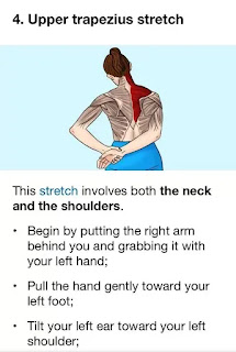
Upper Trapezius Stretch

The following efficient exercises can help reduce pain in the trapezius muscle:

Upper Trapezius Stretch:

  • Keep your back straight and sit or stand tall.
  • Till you feel a stretch along the side of your neck, gently tilt your head to one side and bring your ear towards your shoulder.
  • After holding the stretch for 15 to 30 seconds, move on to the opposite side.
  • Two to three repetitions per side.

Levator Scapulae Stretch:

  • Take a tall stance or sit upright, then slowly turn your head to the side and look over your shoulder.
  • Place your hand on the top of your head and apply gentle pressure to increase the stretch.
  • Hold the stretch for 15-30 seconds, then switch to the other side.
  • Repeat 2-3 times on each side.

Scapular Retraction:

  • Stand with your back against a wall or in a neutral position.
  • As though you were attempting to hold a pencil between your shoulder blades, squeeze them together.
  • Hold the squeeze for 5-10 seconds, then relax.
  • Repeat for 10-15 repetitions.

Shoulder Shrugs:

  • Stand or sit with your arms at your sides.
  • Shrug your shoulders upwards towards your ears as high as possible.
  • Hold for 1-2 seconds, then lower your shoulders back down.
  • Repeat for 10-15 repetitions.

Trapezius Strengthening with Resistance Bands:

  • A stable object at waist height should be used to fasten one end of a resistance band.
  • Hold the other end of the band in one hand and stand with your side facing the anchor point.
  • Keep your arm straight and pull the band diagonally across your body, engaging the trapezius muscle.
  • Slowly return to the starting position.
  • Perform 10-15 repetitions on each side.

Neck Retraction Exercise:

  • Maintain a neutral head posture while sitting or standing.
  • Gently tuck your chin towards your neck, creating a double chin.
  • Hold for a few seconds, then release.
  • Repeat for 10-15 repetitions.

Wall Angels:

  • Stand with your back against a wall and your arms bent at 90 degrees with your elbows and wrists touching the wall.
  • Slowly slide your arms upwards along the wall while keeping your elbows and wrists in contact with the wall.
  • Squeeze your shoulder blades together the entire time you perform the movement.
  • Return your arms to the starting position by sliding them down.
  • Repeat for 10-15 repetitions.

Thoracic Extension Stretch:

  • Sit on the floor with a foam roller positioned horizontally under your upper back.
  • Support your head with your hands, keeping your elbows bent.
  • Slowly lean back over the foam roller, arching your upper back and extending your thoracic spine.
  • After holding the stretch for 15 to 30 seconds, go back to your starting posture.
  • Repeat 2-3 times.

Regularly perform these exercises, paying attention to your form and deliberate movements. Stretch lightly at first, then progressively increase the resistance and intensity as tolerated. Before beginning any new exercise program, especially if you have pre-existing health conditions or concerns, get advice from a medical professional or physical therapist.

Keeping a healthy lifestyle, practicing proper posture, and putting ergonomic techniques into practice can all help prevent pain in the trapezius muscle. 

The following advice can help avoid pain in the trapezius muscle:

Maintain Proper Posture:

Maintain a straight back, relaxed shoulders, and a head that is in line with your spine when you sit or stand.

Stay away from hunching forward or slouching as these postures can strain the trapezius muscle and cause imbalances in the muscles.

Take Regular Breaks:

Take frequent breaks to stand up, stretch, and move around if your job requires you to sit at a desk for extended periods of time or if it's sedentary.

Throughout the day, gently stretch your shoulders and neck to release tension and avoid stiffness.

Practice Ergonomic Work Habits:

To encourage neutral posture and reduce strain on the trapezius muscle, rearrange your workspace.

In order to lessen neck strain, place your computer monitor at eye level and use an ergonomic chair with adequate lumbar support.

Benefit from ergonomic add-ons like supportive cushions, adjustable desks, and ergonomic keyboards.

Use Proper Lifting Techniques:

Use your leg muscles to lift heavy objects instead of your back and shoulders by bending your knees and maintaining a straight back.

When lifting, keep the object close to your body and try not to twist or reach awkwardly.

Stay Active and Exercise Regularly:

Exercise on a regular basis to build strength in the muscles of your neck, shoulders, and upper back, especially the trapezius muscle.

Include activities that help you maintain good posture, like core and back strengthening exercises.

Practice exercises that promote flexibility and mobility, like yoga, Pilates, or swimming.

Manage Stress and Tension:

To release tension in your shoulders and neck, try stress-reduction methods like deep breathing, meditation, or mindfulness.

Throughout the day, take breaks to unwind and reduce stress, particularly if you're dealing with a heavy workload or emotional strain.

Drink plenty of water and eat a balanced diet:

In order to keep muscles hydrated and avoid dehydration, which can exacerbate tension and cramping in the muscles, drink lots of water throughout the day.

To enhance overall muscle health and function, maintain a well-balanced diet high in vitamins, minerals, and nutrients.

Get Adequate Sleep:

Make sure you are getting enough restorative sleep every night by making quality sleep a priority.

Invest in pillows and a supportive mattress to help maintain healthy spinal alignment and lessen shoulder and neck strain when you sleep.

These preventive steps can help lower your chance of experiencing pain in your trapezius muscles and improve your overall musculoskeletal health.

Summary

In summary, pain in the trapezius muscle is a common condition that can greatly affect everyday activities and quality of life. Effective management and prevention of trapezius muscle pain require an understanding of its anatomy, causes, symptoms, and available treatments. The most common causes of pain in the trapezius muscle are injury, stress, poor posture, and overuse of the muscles.

Physiotherapy is essential for reducing pain in the trapezius muscle and enhancing muscle function and mobility. It includes manual therapy techniques, therapeutic exercises, and modalities. Furthermore, preventing trapezius muscle pain can be achieved by putting preventive measures into practice, such as maintaining good posture, adopting ergonomic work habits, being active, controlling stress, and getting enough sleep.

Through the implementation of focused interventions and a comprehensive approach to musculoskeletal health, people can effectively manage pain in the trapezius muscle, enhance their general quality of life, and avoid further episodes of pain. For a customized assessment and treatment recommendations, it's crucial to consult a healthcare provider if you suffer from severe or chronic muscle pain. People may achieve optimal musculoskeletal health and long-term relief through preventive care and lifestyle adjustments.

Saturday, 15 July 2023

Calf Muscle Strain

What is a Calf Muscle Strain?

Calf Muscle strain

A calf muscle strain, also known as a pulled calf muscle, is a common injury that affects the muscles at the back of the lower leg, specifically the two primary calf muscles: the gastrocnemius and the soleus. These muscles play a crucial role in helping you point your toes and push off the ground while walking, running, or jumping.

A calf muscle strain occurs when the muscle fibers are overstretched or torn, usually due to excessive force, sudden movements, or inadequate warm-up before physical activity. Athletes, particularly runners, are more prone to experiencing calf muscle strains, but they can happen to anyone.

Symptoms of a calf muscle strain may include:

Sudden sharp pain in the calf area.

Swelling and tenderness in the affected region.

Limited range of motion in the ankle and foot.

Difficulty walking or bearing weight on the injured leg.

Bruising or discoloration (in more severe cases with extensive muscle tearing).

The severity of a calf muscle strain can vary, ranging from mild to severe. Mild strains may heal on their own with rest, ice, compression, and elevation (RICE), along with avoiding activities that exacerbate the pain. More severe strains may require medical attention, such as physical therapy, stretching exercises, and in rare cases, surgery.

To prevent calf muscle strains, it's essential to properly warm up before engaging in physical activity, stretch regularly, and gradually increase the intensity and duration of exercises. If you experience a calf muscle strain or suspect one, it's best to consult with a healthcare professional for an accurate diagnosis and appropriate treatment plan.

Related Anatomy

To better understand calf muscle strains, let's delve into the related anatomy of the calf region:

Gastrocnemius Muscle: The gastrocnemius muscle is the larger and more superficial of the two calf muscles. It originates just above the back of the knee, with two heads: the medial head and the lateral head. From there, it extends down the back of the lower leg and attaches to the Achilles tendon, which inserts into the heel bone (calcaneus). The gastrocnemius muscle is responsible for flexing the foot at the ankle joint and plays a crucial role in walking, running, and jumping.

Soleus Muscle: The soleus muscle lies deeper beneath the gastrocnemius and is located more towards the midline of the calf. It originates from the upper part of the tibia and fibula, and like the gastrocnemius, it also attaches to the Achilles tendon. The soleus is primarily involved in maintaining an upright posture and helps stabilize the ankle joint during various activities.

Achilles Tendon: The Achilles tendon is a strong, fibrous band that connects the calf muscles (gastrocnemius and soleus) to the heel bone (calcaneus). It is the thickest and strongest tendon in the human body. The Achilles tendon is critical for transmitting the forces generated by the calf muscles to the foot during walking, running, and jumping.

Blood Supply and Nerves: The calf muscles receive their blood supply from branches of the popliteal artery, which is a continuation of the femoral artery in the thigh. Innervation to the calf muscles is provided by the tibial nerve, which is a branch of the sciatic nerve.

Other Structures: In addition to the muscles and tendons, the calf region also contains various blood vessels, nerves, and connective tissues that support the proper functioning of the lower leg.

Injury to any of these structures can lead to a calf muscle strain, where the fibers of the gastrocnemius or soleus muscles get overstretched or torn. Understanding the anatomy of the calf region is crucial for diagnosing and treating calf muscle strains effectively. It also helps in implementing appropriate preventive measures to reduce the risk of such injuries during physical activities.

Type of Calf Muscle Strain

Calf muscle strains are typically categorized based on their severity, with three main types:

  • Grade 1 (Mild): A grade 1 calf muscle strain is the mildest form of strain, where only a few muscle fibers are stretched or slightly torn. Symptoms include mild pain, minimal swelling, and little to no loss of strength or function. The individual may be able to walk with some discomfort. Recovery time for a grade 1 strain is usually relatively short, ranging from a few days to a couple of weeks with proper rest and conservative treatment.
  • Grade 2 (Moderate): A grade 2 calf muscle strain is a more significant injury involving a partial tear of the muscle fibers. The symptoms are more pronounced, with moderate pain, noticeable swelling, and decreased strength and range of motion in the affected leg. Walking becomes difficult, and there might be a visible bruise. Recovery for a grade 2 strain typically takes several weeks, and medical intervention such as physical therapy may be necessary.
  • Grade 3 (Severe): A grade 3 calf muscle strain is the most severe type, involving a complete tear or rupture of the muscle fibers. This can be extremely painful and disabling, causing significant swelling, bruising, and a total loss of function in the affected leg. Walking is usually impossible without assistance. Recovery for a grade 3 strain is more prolonged and may require more aggressive treatment, such as surgery in some cases.

It's crucial to accurately diagnose the grade of the calf muscle strain to determine the appropriate treatment plan. Mild strains can often be managed with rest, ice, compression, and elevation (RICE) along with gentle stretching and exercises as healing progresses. More severe strains might require medical attention, pain management, physical therapy, and a gradual rehabilitation program to restore strength and function.

If you suspect you have a calf muscle strain, especially if the pain is severe or accompanied by significant swelling or bruising, it's essential to seek medical evaluation to determine the extent of the injury and receive appropriate care.

Cause of Calf Muscle Strain

Calf muscle strains are typically caused by the muscles being subjected to excessive force or tension, leading to the stretching or tearing of muscle fibers. Some common causes of calf muscle strains include:

  • Sudden Movements: Abrupt movements, such as jumping or sprinting without proper warm-up, can put a sudden and intense load on the calf muscles, increasing the risk of strain.
  • Overexertion: Engaging in intense physical activity or exercise without adequate conditioning can lead to muscle fatigue and increased susceptibility to strains.
  • Inadequate Warm-up: Failing to warm up before physical activity can leave the muscles unprepared for the stress they are about to experience, making them more susceptible to injury.
  • Muscle Imbalance: Muscle imbalances, where some muscles are stronger than others, can place uneven stress on the calf muscles, making them more prone to strain.
  • Lack of Flexibility: Poor flexibility in the calf muscles and surrounding tissues can lead to limited range of motion and increase the risk of strain during certain movements.
  • Incorrect Footwear: Wearing footwear that does not provide adequate support or has inappropriate shock absorption can contribute to calf muscle strains, especially during running or sports activities.
  • Overtraining or Fatigue: Repeatedly subjecting the calf muscles to intense training without sufficient rest and recovery can weaken the muscles and lead to strains.
  • Age and Previous Injuries: As people age, their muscles may become less flexible and more prone to injury. Additionally, individuals with a history of previous calf muscle injuries may be at a higher risk of re-injury.
  • Direct Trauma: A direct blow or impact to the calf area, such as in contact sports or accidents, can cause a strain in the calf muscles.
  • Poor Biomechanics: Issues with body mechanics or improper form during exercises and activities can put excessive stress on the calf muscles and contribute to strains.

It's essential to listen to your body and gradually build up intensity in physical activities. Engaging in proper warm-up routines, stretching regularly, and using appropriate footwear can help reduce the risk of calf muscle strains. If you do experience a calf muscle strain, giving your muscles adequate time to heal and following a structured rehabilitation program can promote recovery and minimize the chances of future strains.

Symptoms of Calf Muscle Strain

The symptoms of a calf muscle strain can vary depending on the severity of the injury, but common signs and symptoms include:

  • Calf Pain: Sudden, sharp pain in the calf region is the most common and noticeable symptom of a calf muscle strain. The pain may range from mild to severe, depending on the extent of the muscle damage.
  • Swelling: The injured area may become swollen due to inflammation caused by the strain.
  • Tenderness: The calf muscle may feel tender to the touch, and pressing on the affected area can elicit discomfort.
  • Bruising: In more severe strains, bruising (also known as ecchymosis) may appear on the calf due to blood leaking from damaged blood vessels.
  • Limited Range of Motion: The ability to move the ankle and foot might be restricted, especially if the strain affects the gastrocnemius or soleus muscles significantly.
  • Weakness: You may experience weakness in the calf muscles, which can affect your ability to walk or bear weight on the injured leg.
  • Difficulty Walking: Depending on the severity of the strain, walking might become challenging or painful.
  • Muscle Spasms: Some individuals may experience muscle spasms in the calf following the injury.

It's important to note that calf muscle strains are graded based on their severity (as mentioned earlier: grade 1, grade 2, and grade 3). The symptoms can vary accordingly, with grade 1 being the mildest and grade 3 being the most severe.

If you suspect a calf muscle strain, it's essential to avoid putting additional stress on the injured muscle and seek appropriate medical evaluation. A healthcare professional can accurately diagnose the strain's grade and provide appropriate treatment recommendations, which may include rest, ice, compression, elevation (RICE), pain management, and, in more severe cases, physical therapy or other interventions.

Differential Diagnosis

When evaluating a calf muscle injury, healthcare professionals consider various conditions and injuries that can present with similar symptoms to a calf muscle strain. This process is known as the differential diagnosis, where the goal is to rule out other potential causes and arrive at the most accurate diagnosis. Some common conditions that may be included in the differential diagnosis for a calf injury are:

  • Achilles Tendon Rupture: This occurs when the Achilles tendon, which attaches the calf muscles to the heel bone, tears partially or completely. The symptoms can be similar to a severe calf strain, including sudden pain, swelling, bruising, and difficulty walking.
  • Deep Vein Thrombosis (DVT): DVT is a blood clot that forms within the deep veins of the leg, often causing pain, swelling, and warmth in the affected calf. It's crucial to differentiate DVT from a calf strain because DVT can be a potentially serious condition that requires immediate medical attention.
  • Calf Muscle Tear: A calf muscle tear is similar to a calf muscle strain but may involve a more extensive and complete tear of the muscle fibers. The symptoms are generally more severe, and the recovery time might be longer.
  • Muscle Cramps: Sometimes, a simple muscle cramp can cause sudden pain and tightness in the calf. Cramps are usually transient and not associated with swelling or significant tissue damage.
  • Peripheral Arterial Disease (PAD): PAD is a condition where there's reduced blood flow to the lower extremities due to narrowed arteries. It can cause leg pain, especially during physical activity, and can be mistaken for a calf strain.
  • Popliteal Cyst (Baker's Cyst): This is a fluid-filled swelling that can develop at the back of the knee and extend into the calf, causing discomfort and difficulty with knee flexion.
  • Tendinitis: Inflammation of the calf muscle tendons, such as Achilles tendinitis, can cause pain and swelling in the calf area.
  • Nerve Compression or Injury: Conditions like sciatica or tibial nerve compression can cause pain and symptoms in the calf region.

Accurate diagnosis is crucial to ensure appropriate treatment and avoid potential complications. A healthcare professional will typically perform a thorough physical examination, review medical history, and may order imaging tests (such as ultrasound, MRI, or X-rays) to help confirm the diagnosis and rule out other potential causes. If you experience a calf injury or strain, it's essential to seek medical evaluation to receive the appropriate diagnosis and treatment plan.

Diagnosis

The diagnosis of a calf muscle strain typically involves a combination of a thorough physical examination, a review of medical history, and, if necessary, imaging studies. Here's how the diagnosis is typically made:

Medical History: The healthcare professional will begin by asking about the onset of symptoms, the nature of the pain, any preceding activities or trauma that may have triggered the injury, and any relevant medical conditions.

Physical Examination: The healthcare provider will carefully examine the affected calf, looking for signs of swelling, bruising, tenderness, and any other abnormalities. They may also assess the range of motion in the ankle and foot and perform specific tests to assess the strength and integrity of the calf muscles.

Palpation: The doctor will gently feel the calf muscle to identify areas of tenderness and evaluate the extent of the injury.

Functional Assessment: The patient may be asked to perform certain movements, such as walking, standing on tiptoes, and stretching the calf, to determine the level of pain and functional limitations.

Imaging Studies: In some cases, imaging studies may be ordered to confirm the diagnosis and assess the severity of the injury. Common imaging modalities include:

Ultrasound: This can provide real-time images of the calf muscles and tendons, helping to identify muscle tears or other abnormalities.

Magnetic Resonance Imaging (MRI): An MRI can provide detailed images of the calf muscles, tendons, and surrounding structures, helping to visualize any tears or damage more clearly.

X-rays: X-rays are not usually used to diagnose calf muscle strains directly, but they may be done to rule out other potential causes of calf pain, such as bone fractures.

Differential Diagnosis: The healthcare provider will consider other conditions with similar symptoms (as mentioned earlier) to rule out other possible causes of calf pain.

After completing the evaluation, the healthcare professional will determine the grade of the calf muscle strain (if applicable) and provide appropriate treatment recommendations. Treatment may include rest, ice, compression, elevation (RICE), pain management, physical therapy, and a gradual return to activity as the muscle heals.

It's essential to seek medical attention if you suspect a calf muscle strain, especially if the pain is severe, or you have difficulty walking or bearing weight on the injured leg. A proper diagnosis and treatment plan can help facilitate a faster and more complete recovery while minimizing the risk of complications.

Treatment of Calf Muscle Strain

The treatment of a calf muscle strain depends on the severity of the injury, but here are some general guidelines that are commonly followed:

  • Rest: Give your calf muscles adequate rest to allow the injured fibers to heal. Avoid activities that exacerbate the pain or put stress on the calf muscles.
  • Ice: Applying ice to the affected area can help reduce pain and inflammation. Use an ice pack wrapped in a thin cloth and apply it to the calf for about 15-20 minutes every 2-3 hours during the first 48 hours after the injury.
  • Compression: Wearing a compression bandage around the calf can help reduce swelling and provide support to the injured muscles. Be sure not to wrap it too tightly to avoid cutting off circulation.
  • Elevation: Elevating your leg above the level of your heart whenever possible can also help reduce swelling. Prop your leg up on pillows or a cushion while lying down.
  • Pain Management: Over-the-counter pain relievers like ibuprofen or acetaminophen can be used to alleviate pain and reduce inflammation. Always follow the recommended dosage and consult a healthcare professional if you have any health conditions or are taking other medications.
  • Stretching and Gentle Exercises: After the initial acute phase (usually a couple of days), you can begin gentle stretching and range-of-motion exercises to prevent stiffness and improve flexibility. Do these exercises only if they don't cause additional pain.
  • Physical Therapy: In more severe cases or for grade 2 and grade 3 strains, a physical therapist may be involved in the rehabilitation process. They can guide you through a structured program to gradually strengthen and stretch the calf muscles and surrounding tissues.
  • Gradual Return to Activity: As the calf muscles heal and pain subsides, gradually return to normal activities and sports. Avoid rushing back into intense physical activity to prevent reinjury.
  • Supportive Devices: In some cases, your healthcare provider may recommend using crutches or a brace to offload weight from the injured leg and promote healing.
  • Avoiding Risk Factors: Identify and address any contributing factors, such as poor footwear, muscle imbalances, or inadequate warm-up, to reduce the risk of future calf muscle strains.

If the calf muscle strain is severe or not improving with conservative measures, a healthcare professional may consider additional treatments or interventions, such as regenerative therapies, ultrasound-guided injections, or surgery in extreme cases.

It's important to consult with a healthcare provider for an accurate diagnosis and personalized treatment plan based on the specific grade and severity of your calf muscle strain. Proper management and care can help promote healing, reduce complications, and prevent chronic issues.

Physiotherapy Treatment in Calf Muscle Strain

Physiotherapy plays a crucial role in the treatment and rehabilitation of a calf muscle strain. A qualified physiotherapist can design a personalized program to promote healing, reduce pain, restore function, and prevent future injuries. Here are some common physiotherapy interventions used in the treatment of calf muscle strains:

Initial Assessment: The physiotherapist will perform a thorough evaluation to assess the severity of the calf muscle strain, identify any associated factors contributing to the injury, and understand your specific functional limitations and goals.

RICE Protocol: The physiotherapist may initially recommend continuing the Rest, Ice, Compression, and Elevation (RICE) protocol to manage pain and inflammation.

Manual Therapy: Hands-on techniques, such as soft tissue massage, stretching, and mobilization, can help improve blood flow, reduce muscle tightness, and promote healing of the injured calf muscles.

Electrotherapy: Modalities like ultrasound, electrical stimulation, or laser therapy may be used to accelerate tissue healing and reduce pain and inflammation.

Therapeutic Exercises: The physiotherapist will prescribe specific exercises to progressively strengthen the calf muscles, improve flexibility, and enhance functional abilities. Initially, gentle range-of-motion exercises are recommended, gradually progressing to more targeted exercises as the calf heals.

Proprioception and Balance Training: Proprioception exercises help improve the body's awareness of its position in space, enhancing stability and balance. These exercises are essential in preventing reinjury.

Gait and Biomechanical Analysis: The physiotherapist may analyze your walking and running patterns to identify any abnormal biomechanics that could contribute to calf strain. Corrective exercises or gait modifications may be prescribed accordingly.

Modalities for Pain Relief: Apart from ice, the physiotherapist may use other pain-relieving modalities, such as heat therapy or transcutaneous electrical nerve stimulation (TENS), to manage discomfort during the recovery process.

Gradual Return to Activity: The physiotherapist will guide you through a structured plan for safely reintroducing physical activity and sports, taking into account the specific demands of your chosen activities.

Education and Prevention: You will receive education about proper warm-up techniques, stretching exercises, and lifestyle modifications to prevent future calf muscle strains.

Home Exercise Program: To ensure continuity of care, the physiotherapist will provide you with a home exercise program to continue your progress outside of the therapy sessions.

The length of the physiotherapy treatment and the specific techniques used will depend on the severity of the calf muscle strain and the individual's response to therapy. Compliance with the physiotherapy program is essential to achieve optimal results and minimize the risk of reinjury. Always follow your physiotherapist's instructions and communicate any concerns or changes in symptoms during the treatment process.

Rehabilitation of Calf Muscle Strain

Rehabilitation plays a crucial role in the recovery process for a calf muscle strain. The goal of rehabilitation is to promote healing, restore strength and flexibility, improve functional abilities, and prevent future injuries. Here is a general outline of the rehabilitation process for a calf muscle strain:

Rest and Protection: Initially, the injured calf muscle needs rest to allow the damaged fibers to heal. During this period, activities that aggravate the pain should be avoided. Protect the calf from further injury by using crutches or a brace if necessary.

Pain and Inflammation Management: Modalities like ice, heat, and other pain-relieving techniques, as prescribed by your healthcare provider or physiotherapist, can help manage pain and reduce inflammation during the early stages of rehabilitation.

Gentle Range-of-Motion Exercises: Once pain and inflammation have subsided, begin gentle range-of-motion exercises to prevent stiffness and improve flexibility in the calf muscles. An example of a range-of-motion exercise is ankle dorsiflexion and plantar flexion, moving the foot up and down.

Strengthening Exercises: As the calf muscles heal, your physiotherapist will prescribe specific strengthening exercises targeting the gastrocnemius and soleus muscles. These exercises will be gradually progressed based on your tolerance and progress.

Proprioception and Balance Training: Proprioceptive exercises help improve joint stability and body awareness. Incorporate exercises that challenge your balance and coordination, as they play a significant role in preventing future calf muscle strains.

Stretching: Regular stretching of the calf muscles is essential for improving flexibility and preventing muscle imbalances. Your physiotherapist will guide you through appropriate stretching techniques.

Progressive Loading: As your calf muscles gain strength, the rehabilitation program will involve progressive loading through exercises like calf raises, calf presses, and eccentric calf exercises.

Functional Training: Your physiotherapist will tailor exercises to mimic the demands of your daily activities and sports, ensuring a smooth transition back to your regular routines.

Sport-Specific Training: If you're an athlete, the rehabilitation program will include sport-specific exercises to help you safely return to your chosen sport.

Gradual Return to Activity: It's essential to return to physical activity gradually and avoid pushing yourself too hard too soon. Your physiotherapist will guide you through a structured plan for reintroducing activities and sports.

Education and Prevention: Throughout the rehabilitation process, you'll receive education on proper warm-up techniques, injury prevention strategies, and lifestyle modifications to reduce the risk of future calf muscle strains.

Monitoring and Adjustments: Your physiotherapist will continuously monitor your progress and adjust the rehabilitation program as needed to ensure optimal recovery.

Remember that each individual's rehabilitation program is tailored to their specific needs and progress. It's essential to follow your physiotherapist's advice and guidelines to achieve the best possible outcome and reduce the risk of reinjury. Patience and adherence to the rehabilitation plan are key to a successful recovery from a calf muscle strain.

Exercise in Calf Muscle Strain

Exercise plays a critical role in the rehabilitation of a calf muscle strain. However, it's essential to start exercises only after the initial acute phase when pain and inflammation have subsided. Always consult with a healthcare professional or a qualified physiotherapist before starting any exercise program for a calf muscle strain. Below are some examples of calf muscle strain exercises that may be included in a rehabilitation program:

Range-of-Motion Exercises: Gentle ankle movements, such as ankle dorsiflexion (pointing the foot upward) and ankle plantar flexion (pointing the foot downward), can help improve flexibility and prevent stiffness in the calf muscles.

Isometric Exercises: Isometric exercises involve contracting the muscles without changing the joint angle. For the calf muscles, you can perform isometric contractions by pressing your foot into a wall or the floor and holding the contraction for a few seconds. This helps maintain muscle strength without putting excessive stress on the healing fibers.

Calf Raises (Eccentric and Concentric): Calf raises are effective for strengthening the calf muscles. Perform both concentric (raising the heels) and eccentric (lowering the heels) calf raises. For eccentric calf raises, stand on the edge of a step with your heels hanging off the edge, and slowly lower your heels down below the step level, then return to the starting position.

Resistance Band Exercises: Incorporate resistance band exercises for the calf muscles, such as resisted ankle dorsiflexion and plantar flexion.

Toe Tapping: While seated or lying down, gently tap your toes up and down to promote circulation and gentle stretching.

Balance and Proprioception Exercises: Exercises that challenge your balance and improve proprioception (awareness of your body position) can help prevent future injuries. Examples include standing on one leg, single-leg balance exercises on an unstable surface, or using a wobble board.

Heel Walks and Toe Walks: Perform these exercises by walking on your heels and then walking on your toes. These exercises can help strengthen the calf muscles and improve functional abilities.

Calf Stretching: Regularly stretch the calf muscles to improve flexibility. There are various calf stretching exercises, such as the calf wall stretch or using a towel to stretch the calf while sitting.

Incorporate Functional Activities: As you progress, gradually incorporate functional activities into your rehabilitation, such as walking, stair climbing, and controlled jogging.

Remember that the intensity and complexity of exercises should be gradually increased based on your tolerance and progress. If you experience pain or discomfort during any exercise, stop immediately and consult with your healthcare provider or physiotherapist. Adherence to the prescribed exercise program, along with proper rest and recovery, can significantly contribute to the successful rehabilitation of a calf muscle strain and help you return to your regular activities safely.

How long does it take for a pulled calf muscle to heal?

The healing time for a pulled calf muscle, also known as a calf muscle strain, can vary depending on the severity of the injury and the individual's healing capacity. Generally, mild to moderate calf muscle strains (grade 1 and grade 2) may take around 2 to 6 weeks to heal, while more severe strains (grade 3) can take several months to heal completely.

Here's a rough guideline for the healing timeline of a calf muscle strain:

Grade 1 Strain (Mild): Mild calf muscle strains involve minimal tearing of muscle fibers. With appropriate rest, ice, compression, and elevation (RICE), along with gentle stretching and exercises as the muscle heals, most individuals with grade 1 strains can experience significant improvement within 2 to 4 weeks.

Grade 2 Strain (Moderate): Grade 2 strains involve a more significant tear of the muscle fibers. Healing time for grade 2 strains can range from 4 to 6 weeks or more, depending on the extent of the damage and the individual's response to rehabilitation.

Grade 3 Strain (Severe): Severe calf muscle strains, where the muscle fibers are completely torn, may take several months to heal fully. Recovery time can vary, but it's not uncommon for grade 3 strains to require 2 to 3 months or more before significant improvements are seen.

It's essential to note that these timelines are approximate and can vary from person to person. Age, overall health, adherence to the rehabilitation program, and any complications or additional injuries can also influence the healing time.

Throughout the recovery process, it's crucial to follow the advice and guidance of a healthcare professional or a qualified physiotherapist. Trying to rush back into normal activities or sports too soon can increase the risk of reinjury and prolong the healing process. Always listen to your body and allow sufficient time for proper healing before resuming regular physical activities.

Will massage help calf strain?

Massage can be beneficial for a calf strain, but its effectiveness depends on the severity of the strain and the timing of the massage in the healing process. When performed correctly and at the appropriate stage of recovery, massage can help promote healing, reduce muscle tension, improve blood flow, and alleviate pain. Here's how massage can be helpful for a calf strain:

Relaxation and Pain Relief: Massage can help relax tense muscles and release muscle spasms, which can provide pain relief and improve overall comfort during the healing process.

Improved Circulation: Massage increases blood flow to the injured area, which can enhance the delivery of oxygen and nutrients to the healing tissues, promoting faster recovery.

Reduced Scar Tissue Formation: Gentle massage techniques may help reduce the formation of excessive scar tissue, which can impede proper muscle healing and lead to long-term complications.

Increased Flexibility: As the healing progresses, massage can help improve flexibility and range of motion in the calf muscles.

However, it's essential to use caution with massage in the early stages of a calf strain, especially in the acute phase when the injury is fresh and painful. In the initial days after the injury, the calf muscle may be inflamed and tender, making deep massage or direct pressure on the affected area uncomfortable or even harmful. In this acute phase, other therapies like rest, ice, compression, and elevation (RICE) are more appropriate.

Before seeking a massage for a calf strain, it's best to consult with a qualified healthcare professional or a licensed massage therapist who is experienced in dealing with sports injuries. They can assess the severity of the strain, consider any potential contraindications, and recommend the appropriate timing and techniques for massage.

As the calf strain progresses through the healing stages and pain and inflammation subside, gentle massage, stretching, and other soft tissue techniques can be incorporated into the rehabilitation process to support recovery and enhance the overall healing process.

How to Prevent Calf Muscle Strain?

To prevent calf muscle strains, you can follow these preventive measures:

Proper Warm-up: Always warm up before engaging in physical activities or exercise. Include dynamic stretching and light aerobic exercises to increase blood flow and prepare your calf muscles for the upcoming demands.

Gradual Progression: Avoid sudden increases in the intensity, duration, or frequency of your workouts. Gradually progress your exercises to allow your calf muscles to adapt and strengthen over time.

 Stretching: Incorporate regular calf stretches into your routine to maintain flexibility in the calf muscles. Focus on both gastrocnemius and soleus stretches to improve overall calf flexibility.

Strength Training: Include calf-strengthening exercises in your workout routine. Focus on exercises like calf raises and toe raises to build strength in the calf muscles.

Balanced Footwear: Wear appropriate and well-fitting footwear for your activities. Choose shoes that provide adequate support and cushioning, especially for high-impact activities like running or jumping.

Proper Foot Biomechanics: Consider getting your foot mechanics evaluated by a professional, such as a podiatrist or physiotherapist. Address any foot imbalances or abnormalities that may contribute to calf strain.

Hydration: Stay well-hydrated during physical activities, as dehydration can increase the risk of muscle cramps and strains.

Rest and Recovery: Allow sufficient time for rest and recovery between workouts to avoid overuse injuries and give your calf muscles time to repair and strengthen.

Cross-Training: Incorporate cross-training activities into your routine to avoid overloading the calf muscles with repetitive movements. Varying your workouts can help prevent strain from repetitive stress.

Proper Technique: Pay attention to proper form and technique during exercises and sports activities. Seek guidance from a qualified trainer or coach if needed.

Warm-down and Stretching: After intense physical activity, perform a cool-down routine, followed by static stretching, to promote muscle relaxation and flexibility.

Listen to Your Body: Pay attention to any signs of discomfort or pain in the calf region. If you feel any discomfort, take a break from the activity and allow your calf muscles to rest and recover.

By incorporating these preventive measures into your fitness routine and being mindful of your calf muscles' well-being, you can significantly reduce the risk of calf muscle strains and other related injuries. If you have a history of calf strains or any concerns, consider consulting with a healthcare professional or a qualified trainer to design a personalized prevention plan.

Conclusion

In conclusion, a calf muscle strain is a common injury characterized by the stretching or tearing of the calf muscle fibers. It can occur due to sudden movements, overexertion, inadequate warm-up, muscle imbalances, or direct trauma. Understanding the related anatomy of the calf region is essential for accurate diagnosis and treatment.

Calf muscle strains are typically categorized into three grades based on their severity, with grade 1 being mild, grade 2 being moderate, and grade 3 being severe. The symptoms of a calf muscle strain include pain, swelling, tenderness, bruising, limited range of motion, weakness, and difficulty walking.

The differential diagnosis involves considering other conditions with similar symptoms to rule out alternative causes. Once diagnosed, the treatment of a calf muscle strain involves the RICE protocol (rest, ice, compression, and elevation), pain management, physical therapy, and a gradual return to activity.

The rehabilitation process focuses on promoting healing, restoring strength and flexibility, and preventing future injuries. Physiotherapy plays a crucial role in designing personalized exercise programs and guiding patients through a structured plan.

Prevention is key to avoiding calf muscle strains. Incorporating proper warm-up, gradual progression, stretching, strengthening, balanced footwear, and listening to your body are essential preventive measures. Regularly seeking professional guidance and paying attention to biomechanics can further reduce the risk of calf muscle strains.

Overall, understanding the causes, symptoms, diagnosis, treatment, and prevention of calf muscle strains empowers individuals to manage these injuries effectively and supports a safe and successful recovery process.

Saturday, 15 April 2023

Peroneal tendon tear: Cause, Symptoms, Treatment, Exercise

What is a Peroneal tendon tear?

Peroneal tendon tear

Peroneal tendon tear is a condition where one or both tendons that run down the outer side of the lower leg, behind the ankle and into the foot, are partially or completely torn. These tendons are responsible for stabilizing the ankle and foot during movement, especially during activities that involve twisting or turning of the foot. 

A tear in the peroneal tendons can lead to pain, weakness, swelling, and instability in the ankle and foot. It can be caused by repetitive strain, overuse, or traumatic injury to the tendons. Treatment for peroneal tendon tear may involve rest, immobilization, physical therapy, or surgery depending on the severity of the injury.

Related Anatomy

The peroneal tendons are two fibrous cords that originate from the muscles in the outer part of the lower leg, the peroneus longus, and peroneus brevis muscles. The peroneus longus tendon runs under the foot and attaches to the first metatarsal bone, while the peroneus brevis tendon runs behind the ankle and attaches to the fifth metatarsal bone. 

These tendons work together to stabilize the ankle and foot during movement, especially during activities that involve twisting or turning of the foot. The tendons are surrounded by a protective sheath and held in place by ligaments and other soft tissues. The blood supply to the peroneal tendons is limited, which can make them susceptible to injury and slow to heal.

Causes of Peroneal tendon tear

Peroneal tendon tears can be caused by a variety of factors, including:

  • Repetitive Strain: Activities that involve repetitive motions, such as running, jumping, and dancing, can put a strain on the peroneal tendons and lead to small tears over time.
  • Ankle Sprains: A severe ankle sprain or twisting injury can cause a peroneal tendon tear.
  • Overuse: Overuse of the peroneal tendons without adequate rest and recovery can lead to tendon damage and eventually tears.
  • Degeneration: Aging and wear and tear can lead to degeneration of the peroneal tendons, making them more prone to injury.
  • Abnormal Foot Mechanics: Certain foot deformities, such as flat feet or high arches, can place extra stress on the peroneal tendons and increase the risk of tears.
  • Trauma: Direct trauma to the ankle or lower leg, such as a fall or impact injury, can cause a peroneal tendon tear.
  • Improper Footwear: Wearing shoes that do not fit properly or provide adequate support can increase the risk of peroneal tendon injuries.

Symptoms of Peroneal tendon tear

The symptoms of a peroneal tendon tear can vary depending on the severity of the injury. Common symptoms include:

  • Pain: The most common symptom of a peroneal tendon tear is pain on the outside of the ankle and foot, especially when bearing weight or moving the foot.
  • Swelling: The affected area may be swollen and tender to the touch.
  • Weakness: A peroneal tendon tear can cause weakness in the ankle and foot, making it difficult to move the foot or maintain balance.
  • Instability: The ankle may feel unstable or give way, especially when walking on uneven surfaces.
  • Clicking or popping: A popping or clicking sound may be heard or felt when moving the ankle or foot.
  • Redness or warmth: The skin around the affected area may be red or warm to the touch.
  • Limited range of motion: A peroneal tendon tear can limit the range of motion of the ankle and foot, making it difficult to perform activities such as walking, running, or jumping.

It is important to seek medical attention if you experience any of these symptoms, as untreated peroneal tendon tears can lead to chronic ankle instability and other complications.

Risk Factor

Several risk factors may increase the likelihood of developing a peroneal tendon tear, including:

  • Age: Tendon degeneration and wear and tear increases with age, making older individuals more susceptible to tendon injuries.
  • Overuse: Repetitive activities that place stress on the peroneal tendons, such as running or jumping, increase the risk of developing a tear.
  • Improper Footwear: Wearing shoes that do not fit properly or provide adequate support can increase the risk of developing a peroneal tendon tear.
  • Foot Deformities: Certain foot deformities, such as flat feet or high arches, can put extra stress on the peroneal tendons and increase the risk of developing a tear.
  • Trauma: A traumatic injury, such as a fall or ankle sprain, can cause a peroneal tendon tear.
  • Sports Participation: Participation in sports that involve twisting or turning of the foot, such as soccer or basketball, can increase the risk of developing a peroneal tendon tear.
  • Weakness or Tightness in Muscles: Weakness or tightness in the muscles surrounding the ankle and foot can affect the alignment of the foot, putting additional stress on the peroneal tendons.

It is important to be aware of these risk factors and take steps to prevent peroneal tendon tears, such as wearing appropriate footwear, using proper technique during physical activity, and maintaining strong and flexible muscles in the lower leg and foot.

Differential Diagnosis

The symptoms of a peroneal tendon tear can be similar to those of other conditions affecting the ankle and foot. Therefore, it is important to consider other possible diagnoses when evaluating a patient with these symptoms. Some of the differential diagnoses that may need to be ruled out include:

  • Ankle sprain: An ankle sprain is a common injury that can cause pain, swelling, and instability in the ankle. It occurs when the ligaments that support the ankle joint are stretched or torn.
  • Achilles tendonitis: Achilles tendonitis is an overuse injury that affects the Achilles tendon, which connects the calf muscles to the heel bone. It can cause pain, swelling, and stiffness in the back of the ankle and lower leg.
  • Stress fracture: A stress fracture is a small crack in a bone that can occur due to overuse or repetitive stress. It can cause pain, swelling, and tenderness in the affected area.
  • Plantar fasciitis: Plantar fasciitis is a condition that affects the plantar fascia, a thick band of tissue that runs along the bottom of the foot. It can cause heel pain, especially in the morning or after prolonged periods of standing or walking.
  • Tarsal tunnel syndrome: Tarsal tunnel syndrome is a condition that occurs when the posterior tibial nerve, which runs through a narrow passage in the ankle, becomes compressed. It can cause pain, numbness, and tingling in the ankle and foot.
  • Arthritis: Arthritis is a condition that causes inflammation in the joints, which can lead to pain, swelling, and stiffness. It can affect any joint in the body, including the ankle and foot.

It is important to consult a healthcare professional for an accurate diagnosis, as treatment can vary depending on the underlying cause of the symptoms.

Diagnosis

The diagnosis of a peroneal tendon tear typically begins with a physical examination by a healthcare professional. During the examination, the healthcare professional will look for signs of swelling, tenderness, instability, and weakness in the ankle and foot. They may also ask about the patient's medical history, including any previous injuries or conditions that may affect the ankle or foot.

If a peroneal tendon tear is suspected, imaging tests may be ordered to confirm the diagnosis and determine the severity of the injury. Common imaging tests used to diagnose a peroneal tendon tear include:

  • X-ray: An X-ray may be ordered to rule out the possibility of a bone fracture or other bony abnormalities.
  • Magnetic resonance imaging (MRI): An MRI uses a magnetic field and radio waves to produce detailed images of the soft tissues in the ankle and foot, including the peroneal tendons. It can help to identify the location and severity of a peroneal tendon tear.
  • Ultrasound: An ultrasound uses high-frequency sound waves to produce images of the soft tissues in the ankle and foot, including the peroneal tendons. It can help to identify the presence and location of a tear.

Once a diagnosis is confirmed, the healthcare professional will develop a treatment plan based on the severity of the injury and the patient's individual needs and goals. Treatment may include rest, ice, compression, elevation, physical therapy, and in some cases, surgery.

Peroneal tendon tear Test

There are several tests that can be performed to help diagnose a peroneal tendon tear. These tests are usually performed by a healthcare professional, such as a doctor or physical therapist. Some common tests include:

  • Palpation: The healthcare professional may gently press and feel along the path of the peroneal tendons to check for tenderness or swelling.
  • Range of motion: The healthcare professional may check the range of motion of the ankle and foot, looking for any limitations or pain during movement.
  • Resistance tests: The healthcare professional may apply resistance to the ankle and foot to check for weakness or pain.
  • Functional tests: The healthcare professional may ask the patient to perform specific movements, such as standing on the affected foot or walking on their toes or heels, to assess function and stability.
  • Imaging tests: Imaging tests, such as X-rays or MRI, may be ordered to provide more detailed information about the extent of the injury.

It is important to seek prompt medical attention if you suspect a peroneal tendon tear, as an accurate diagnosis is key to developing an effective treatment plan.

Treatment of Peroneal tendon tear

The treatment for a peroneal tendon tear will depend on the severity of the injury. In general, treatment options may include:

  • Rest and immobilization: The first step in treating a peroneal tendon tear is to rest and protect the affected foot and ankle. A healthcare professional may recommend immobilization with a cast, brace, or walking boot to limit movement and allow the tendon to heal.
  • Ice and compression: Applying ice and compression to the affected area can help to reduce swelling, inflammation, and pain.
  • Physical therapy: Physical therapy exercises can help to strengthen the muscles and improve the range of motion in the ankle and foot. This may involve stretching, range of motion exercises, and strengthening exercises.
  • Medications: Over-the-counter pain medications, such as acetaminophen or ibuprofen, may be recommended to help manage pain and inflammation.
  • Corticosteroid injections: In some cases, a healthcare professional may recommend a corticosteroid injection to help reduce inflammation and pain.
  • Surgery: If the tear is severe or does not respond to non-surgical treatments, surgery may be necessary. Surgery may involve repairing the torn tendon or, in some cases, transferring a nearby tendon to replace the damaged peroneal tendon.
  • Orthotics: Wearing a supportive brace or custom orthotics can help to provide additional support and prevent further injury.

It is important to follow a healthcare professional's recommendations for treatment and allow sufficient time for the tendon to heal before returning to physical activity. With proper treatment and rehabilitation, most people with a peroneal tendon tear can return to their normal activities.

Physical Therapy Treatment

Physical therapy can be an important part of the treatment plan for a peroneal tendon tear. The goals of physical therapy are to improve range of motion, strength, and function of the ankle and foot, and to prevent future injury. Physical therapy treatment may include:

  • Range of motion exercises: These exercises help to improve the flexibility and mobility of the ankle and foot, which can help to prevent stiffness and promote healing.
  • Strengthening exercises: Strengthening exercises can help to improve the strength and stability of the ankle and foot. This may involve exercises to strengthen the peroneal muscles, as well as other muscles in the ankle and foot.
  • Balance and proprioceptive training: Balance and proprioceptive exercises can help to improve the body's ability to sense and respond to changes in position and movement, which can reduce the risk of future injury.
  • Gait training: Gait training can help to improve walking and running mechanics, which can help to reduce stress on the peroneal tendons and promote healing.
  • Modalities: Physical therapists may use various modalities such as ultrasound, electrical stimulation, and heat or ice therapy to help reduce pain and inflammation.
  • Education: Physical therapists may provide education on proper footwear, exercise techniques, and strategies to prevent future injury.

The duration of physical therapy treatment will depend on the severity of the injury and the individual's response to treatment. It is important to work closely with a physical therapist to ensure that exercises are performed correctly and safely, and to progress through the rehabilitation program at an appropriate pace.

Complications of Peroneal tendon tear

Complications of a peroneal tendon tear can arise if the injury is not properly treated or if the patient returns to physical activity too soon. Some of the possible complications include:

  • Chronic pain: If the peroneal tendon tear does not heal properly, it can lead to chronic pain and discomfort in the ankle and foot.
  • Recurrent injuries: If the peroneal tendons are weakened or damaged, they may be more susceptible to future injuries, especially if the patient returns to physical activity too soon.
  • Ankle instability: Damage to the peroneal tendons can also lead to ankle instability, which can increase the risk of ankle sprains and other injuries.
  • Tendon rupture: In some cases, a severe peroneal tendon tear can lead to a complete tendon rupture, which may require surgical repair.
  • Posterior tibial tendon dysfunction: The peroneal tendons work in conjunction with other tendons and muscles in the foot and ankle. Damage to the peroneal tendons can affect the function of other structures in the foot, such as the posterior tibial tendon, leading to additional complications.

It is important to seek prompt medical attention and follow a healthcare professional's recommendations for treatment and rehabilitation to minimize the risk of complications.

How to Prevent Peroneal tendon tear?

There are several steps that can be taken to help prevent a peroneal tendon tear:

  • Wear appropriate footwear: Wearing shoes that provide good support and fit properly can help to reduce the risk of ankle and foot injuries, including peroneal tendon tears.
  • Warm-up and stretch: Engaging in a proper warm-up and stretching routine before physical activity can help to prepare the muscles and tendons for movement and reduce the risk of injury.
  • Gradually increase activity level: Gradually increasing the intensity and duration of physical activity can help to prevent overuse injuries, including peroneal tendon tears.
  • Strengthen the ankle and foot muscles: Strong muscles in the ankle and foot can help to provide support and stability to the tendons and reduce the risk of injury.
  • Address underlying conditions: Certain conditions, such as flat feet or high arches, can increase the risk of peroneal tendon tears. Addressing these underlying conditions, such as with custom orthotics or physical therapy, can help to reduce the risk of injury.
  • Rest and recovery: Allowing sufficient time for rest and recovery between physical activity sessions can help to prevent overuse injuries, including peroneal tendon tears.
  • Seek prompt medical attention: If you experience symptoms of a peroneal tendon tear or any other ankle or foot injury, seek prompt medical attention to receive an accurate diagnosis and appropriate treatment.

By following these steps, individuals can help to reduce the risk of a peroneal tendon tear and other ankle and foot injuries.

Can Peroneal tendon tear heal without surgery?

In many cases, a peroneal tendon tear can heal without surgery, particularly if it is a partial tear or if the tear is detected and treated early. However, the specific treatment plan will depend on the severity and extent of the tear, as well as other factors such as the patient's age, overall health, and activity level.

Conservative treatment options for a peroneal tendon tear may include rest, ice, compression, elevation, and physical therapy to help improve strength, flexibility, and range of motion. In some cases, a brace or cast may be recommended to immobilize the ankle and foot and allow the tendon to heal.

Surgery may be necessary for more severe or complex peroneal tendon tears, such as complete tears or tears that involve multiple tendons. Surgery may also be recommended if conservative treatments have not been effective in improving symptoms or if the tear is causing significant instability or dysfunction of the ankle and foot.

It is important to work closely with a healthcare professional to determine the most appropriate treatment plan for a peroneal tendon tear. Delaying treatment or failing to properly address the injury can increase the risk of complications and long-term problems.

Is Peroneal tendon surgery worth it?

Whether or not peroneal tendon surgery is worth it depends on several factors, including the severity of the injury, the patient's overall health and activity level, and the potential risks and benefits of the surgery.

In some cases, surgery may be necessary to repair a complete or complex peroneal tendon tear or to address other structural problems in the ankle or foot that are contributing to the injury. Surgery may also be recommended if conservative treatments have not been effective in improving symptoms or if the tear is causing significant instability or dysfunction of the ankle and foot.

However, surgery also carries risks, including the potential for infection, nerve damage, or other complications. Recovery from surgery can also be a lengthy process, and may require a period of immobilization, physical therapy, and activity restrictions.

Ultimately, the decision to undergo peroneal tendon surgery should be made in consultation with a healthcare professional, who can help weigh the potential risks and benefits of the procedure and determine the most appropriate treatment plan based on the individual patient's needs and circumstances.

Can a peroneal tendon tear heal on its own?

In some cases, a peroneal tendon tear can heal on its own, particularly if it is a partial tear or if the tear is detected and treated early. However, the specific treatment plan will depend on the severity and extent of the tear, as well as other factors such as the patient's age, overall health, and activity level.

Conservative treatment options for a peroneal tendon tear may include rest, ice, compression, elevation, and physical therapy to help improve strength, flexibility, and range of motion. In some cases, a brace or cast may be recommended to immobilize the ankle and foot and allow the tendon to heal.

However, complete tears of the peroneal tendon or more severe injuries may not heal on their own and may require surgical intervention to repair the tendon.

It is important to work closely with a healthcare professional to determine the most appropriate treatment plan for a peroneal tendon tear. Delaying treatment or failing to properly address the injury can increase the risk of complications and long-term problems.

How long does it take to heal a torn peroneal tendon?

The length of time it takes to heal a torn peroneal tendon can vary depending on the severity and extent of the tear, as well as the specific treatment approach used. In general, it can take several weeks to several months to fully heal a torn peroneal tendon.

For minor or partial tears, conservative treatment options such as rest, ice, compression, and physical therapy may be effective in promoting healing and reducing symptoms. With these treatments, patients may begin to see improvement in their symptoms within a few weeks and may be able to gradually return to their normal activities over the course of several months.

For more severe or complete tears, surgery may be necessary to repair the tendon. Following surgery, patients may need to wear a cast or brace for several weeks to allow the tendon to heal. Physical therapy and rehabilitation exercises will also be necessary to help rebuild strength and restore range of motion.

It is important for patients to follow their healthcare professional's recommended treatment plan and allow for sufficient healing time to avoid reinjury or other complications.

Summary

A peroneal tendon tear is a type of ankle injury that can be caused by a sudden trauma or overuse. The peroneal tendons run along the outside of the ankle and help to stabilize the ankle and foot. Symptoms of a peroneal tendon tear may include pain, swelling, weakness, and difficulty with walking or running. Treatment for a peroneal tendon tear may include rest, ice, compression, physical therapy, and in some cases, surgery. 

Physical therapy can be an important part of the treatment plan, helping to improve range of motion, strength, and function of the ankle and foot, and to prevent future injury. Complications of a peroneal tendon tear can arise if the injury is not properly treated, including chronic pain, recurrent injuries, ankle instability, tendon rupture, and dysfunction of other structures in the foot. To prevent a peroneal tendon tear, individuals should wear appropriate footwear, warm-up and stretch, gradually increase activity level, strengthen the ankle and foot muscles, address underlying conditions, rest and recover, and seek prompt medical attention.