Showing posts with label Anatomy. Show all posts

Thursday, 14 November 2024

Femur Bone Anatomy

Introduction

Femur Bone Anatomy
Femur Bone Anatomy

The femur, commonly known as the thigh bone, is the longest, strongest, and heaviest bone in the human body. Located in the upper leg, it extends from the hip joint down to the knee joint, playing a crucial role in supporting the body’s weight and enabling a wide range of movements, including walking, running, and jumping.

Its unique structure provides leverage and stability, with the femoral head fitting into the hip socket to form a ball-and-socket joint, allowing for a smooth, multi-directional movement. Due to its weight-bearing function, the femur is particularly resilient but can be vulnerable to fractures, especially in high-impact injuries or conditions like osteoporosis.

Structure of Femur Bone

The proximal ends of the tibiae articulate with the two femurs as they converge medially toward the knees. The femora of female humans converge more than those of men due to the thickness of their pelvic bones. The femurs converge so far that the knees contact in genu valgum, also known as knock knee.

Upper part

The head, neck, two trochanters, and surrounding components are located at the upper or proximal extremity, which is near the torso. The femoral extremities are thickest in the lower extremity and thinnest in the higher extremity.

The neck is squeezed in the middle and measures 4 to 5 cm in length, with the lowest diameter from front to rear. It is around 150 degrees in a baby and, on average, drops to 120 degrees in old age. The head of the femur can be felt deep as a resistance profound (deep) for the femoral artery in thin individuals with the thigh laterally turned.

Both the bigger and minor trochanters may be found here. The femur’s most lateral noticeable part is the greater trochanter, which has an almost box-like form. It’s easy to feel the larger trochanter. The lowest portion of the femur neck extends in a cone form as the lesser trochanter.

The linea quadrata, also known as the quadrate line, is a faint ridge that occasionally appears down the back of the body, starting at the center of the intertrochanteric crest.

The quadrate tubercle is situated on the intertrochanteric crest, about where the upper one-third and lower two-thirds meet. The epiphyseal line runs straight through the quadrate tubercle, according to a brief anatomical research.

Body of Femur Bone

The linea aspera, a conspicuous longitudinal ridge that diverges proximally and distally as the medial and lateral ridges, strengthens it. It is somewhat arched, making it convex in front and concave behind. The shaft cannot be felt because of the thigh’s extensive musculature.

Its occurrence varies amongst ethnic groups, ranging from 17 to 72%, and it is often reported that females are more likely than men to have it.

Lower part

Although its transverse diameter is larger than its anteroposteriorly (front to back) diameter, it has a slightly cuboid shape. It is made up of two condyles, which are oblong protuberances.

The patellar surface is a smooth, shallow articular depression that separates the somewhat protruding condyles on the anterior side. They protrude significantly posteriorly, and between them is a deep indentation known as the femur’s intercondylar fossa. The condyles are not quite parallel to one another; the medial’s long axis travels rearward and medially, whereas the lateral’s is almost immediately anteroposterior. The anterior cruciate ligament of the knee joint is related to an impression on the higher and rear portions of its lateral wall, while the posterior cruciate ligament.

It has two convexities, the lateral of which is wider, more noticeable, and reaches higher than the medial, as well as a median groove that runs downhill to the intercondyloid fossa.

Development

The ectoderm and the underlying mesoderm interact to produce the femur from the limb buds; this process takes place approximately during the fourth week of development.

By the conclusion of the embryonic phase, endochondral ossification has started, and by the 12th week of development, all of the long bones of the limbs, including the femur, have main ossification centers.

Blood Supply

Once the artery has passed through the ilioinguinal ligament, it becomes the main branch of the external iliac artery. A branch of the femoral artery is the medial and lateral circumflex arteries. They supply the femoral head through important anastomotic connections, as does the obturator artery, a branch of the internal iliac artery. The deep femoral artery’s perforating branches provide blood to the femur’s shaft and distal region.

Function of Femur Bone

The femur is also the origin of several biarticular muscles, such as the gastrocnemius and plantaris muscles, which bridge two joints. There are a total of 23 distinct muscles that either attach to or arise from the femur. The thigh is shown in cross-section as having three distinct fascial compartments, each of which has muscles.

Embryology

Leg growth and development are induced by the limb bud’s apical ectodermal ridge. The femur develops from endochondral ossification, in which bone replaces hyaline cartilage models, and is derived from the lateral plate somatic mesoderm of the lower limb bud. There is no cartilage model for the intramembranous ossification process that creates articular cartilages and epiphyseal plates. It is the myotomic component of the somites that produces the femur muscles. The periosteum, which envelops the femur, provides nourishment via the nearby blood supply.

Clinical significance

Fractures

Particularly when osteoporosis is present, a femur fracture that affects the femoral head, femoral neck, or the shaft of the femur just below the lesser trochanter may be categorized as a hip fracture. A traction splint can be used to treat femur fractures in a pre-hospital environment.

Adolescent Hip Disorder

Overweight teenage boys are more likely to have slipped capital femoral epiphysis (SCFE), a hip condition affecting the femoral head. Although the reason is frequently idiopathic, radiation treatment, renal failure, and endocrine dysfunction have all been linked to SCFE. Although early treatment with a single screw through the growth plate has been demonstrated to prevent increasing slippage, preventative therapy for SCFE remains debatable.

The modified Dunn method entails fixing the femoral head with screws after removing a wedge of the femoral neck to rectify the deformity. With the modified Dunn surgery, avascular necrosis and persistent hip pain were frequent post-operative sequelae.

Vascular

When the blood flow to the femoral head is interrupted, an uncommon pediatric condition known as Legg-Calve-Perthes disease (LCP) results. Twin research indicates that environmental variables including low socioeconomic status increase the likelihood of LCP. Inguinal hernias, Down syndrome, and genitourinary disorders are among the congenital deformities linked to LCP. The child’s age and illness stage determine how they should be managed. LCP can be treated with braces, acupuncture, exercise, bisphosphonates, and hip arthroscopy.

Environmental

A typical malformation of the long bones is caused by rickets, which is an inability to mineralize bone. The main mechanism by which the growth plate calcifies and encourages the formation of long bones is endochondral ossification. However, rickets reduce or eliminate this process. Atypical phosphate metabolism, decreased sun exposure, and inadequate calcium or phosphate consumption are further factors.

  • Injuries and Conditions
  • Femoral fractures
  • Femoral Stress fracture
  • Patellofemoral pain syndrome
  • Femoral Neck Fracture
  • Greater Trochanteric Pain Syndrome

Wednesday, 6 March 2024

Bicipital Groove

Bicipital Groove
Bicipital Groove

The humerus bone of the upper arm contains an anatomical feature called the Bicipital Groove, which is also referred to as the intertubercular groove or the sulcus. The long head of the biceps brachii muscle tendon, which extends from the shoulder to the elbow, is accommodated and protected by the bicipital groove, which is located on the anterior (front) aspect of the bone.

It permits the long tendon of the biceps brachii muscle to pass through. This unique groove facilitates the smooth movement of the arm and efficient transfer of muscular forces by supporting the stability and healthy operation of the biceps tendon.

Studying human anatomy requires an understanding of the structure and function of the bicipital groove, especially in order to understand upper limb mechanics and the relationships between bones, muscles, and tendons.

The bicipital groove splits the greater and smaller tubercles. Adults are about 8 cm long, 4–6 mm deep, and 1 cm wide on average. The long biceps brachii tendon lodges between the pectoralis major tendons on the lateral lip and the teres major tendons on the medial lip. An additional branch of the anterior humeral circumflex artery originates at the shoulder joint.

The latissimus dorsi muscle inserts into the bicipital groove floor. The teres major muscle inserts into the groove on the medial lip.

It ends near the location where the top part of the bone joins the middle third, curving downward.  It is the axilla's lateral wall.

Attachments of Bicipital Groove

Musculotendinous

Three tendons attach to the bicipital groove are:

  • lateral lip: pectoralis major
  • floor: latissimus dorsi
  • medial lip: teres major

Relations and/or Boundaries

The bicipital groove is located on the anterior surface of the proximal humerus and has the following boundaries:

  • Superiorly: transverse humeral ligament
  • Laterally: greater tuberosity of the humerus
  • Medially: lesser tuberosity of the humerus

Function

The long tendon of the biceps brachii muscle can pass through the bicipital groove.

The transverse humeral ligament and the muscle fibers that extend from it stabilize and facilitate the tendon of the long head of the biceps brachi muscle in this groove, preventing subluxation during multidirectional biomechanical movements of the arms. Furthermore, the main biomechanical actions of the biceps brachi muscle, whose tendon is situated in the bicipital groove, are supination, flexion, and screwing.

Clinical Importance

  • Long head of biceps tendon dislocation
  • Bicipital Tendinitis
  • Pectoralis major tear
  • Latissimus dorsi tear

Friday, 16 February 2024

Subscapularis Muscle

What is the Subscapularis Muscle?
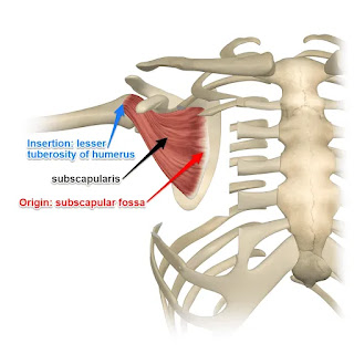
Subscapularis Muscle

The subscapularis is a broad, triangular muscle that inserts into the front of the shoulder-joint capsule, the lesser tubercle of the humerus, and the subscapular fossa.

It is one of the four rotator cuff muscles, along with the teres minor, infraspinatus, and supraspinatus. The biggest and strongest rotator cuff muscle is the subscapularis.

Weakness in internal rotation results from function loss caused by injury to the muscle or tendon. The preferred course of therapy for tendinopathies and partial tears is non-operative care.

If conservative therapy is unsuccessful, (1) surgical surgery may be necessary; (2) based on the individual’s activity level or occupation (athletes, for example); and (3) in the event of full-thickness rips.

Structure

A thick fascia covering the subscapularis joins to the scapula at the borders of the attachment (origin) of the subscapularis.

The fibers of the muscle merge into an insertion tendon after passing laterally from the muscle’s origin. The glenohumeral (shoulder) joint capsule and the tendon are intermingled.

A bursa sits between the tendon and a bare spot at the lateral angle of the scapula the neck of the scapula. It is connected to the cavity of the shoulder joint by an opening in the joint capsule. The serratus anterior and subscapularis are divided by the subscapularis (supra serratus) bursa.

Origin of Subscapularis muscle

It originates from the bottom two-thirds of the groove on the axillary border (subscapular fossa) of the scapula, the intermuscular septa (which form ridges on the scapula), and the medial two-thirds of the scapula's costal surface.

Aponeurosis, which divides the muscle from the teres major and the long head of the triceps brachii, is the source of some fibers. Tendinous laminae, which cross the muscle and are linked to ridges on the bone, are the source of other fibers.

Insertion of Subscapularis muscle

It attaches to the anterior portion of the shoulder joint capsule as well as the smaller tubercle of the humerus. Tendinous fibers penetrate the bicipital groove and extend to the larger tubercle.

Nerve Supply

The posterior cord of the brachial plexus (C5 to C7) contains branches that give rise to the subscapular nerve, which trifurcates into the upper, middle, and lower subscapular nerves. Both the upper and lower subscapular nerves supply the subscapularis.

The cranial half of the muscle is innervated by the upper subscapular nerve, whereas the caudal half of the muscle is innervated by the lower subscapular nerve, which bifurcates into two branches.

The latissimus dorsi muscle is innervated by the middle scapular nerve, also referred to as the thoracodorsal nerve, whereas the teres major is innervated by the inferior branch of the lower subscapular nerve.

Blood Supply and Lymphatics

The main blood supply to the subscapularis muscle is provided by the subscapular artery, which is a branch of the axillary artery. The lymph nodes in the axilla receive lymph drainage.

Course of the Muscles

The subscapularis muscle enters into the humerus’s lesser tubercle after starting at the subscapular fossa. The muscle adducts the humerus and spins it inside out. In the bicipital groove, the bicep tendon is located beneath the subscapularis tendon.

Innervation

The upper and lower subscapular nerves (C5–C6), which are branches of the posterior chord of the brachial plexus, supply the subscapularis.

Actions/movements

The humerus’s internal rotation is its primary function. In specific postures, it aids with shoulder adduction and extension.

The actions of this muscle are significantly influenced by the position of the arm: when the arm is raised, the subscapularis pulls the humerus forward and downward; when the humerus is fixed, the insertion of the subscapularis can act as an origin and cause the inferior border of the scapula to abduct.

Function of Subscapularis muscle

As the proximal humerus is fixed during elbow, wrist, and hand motions, the subscapularis helps to stabilize the shoulder joint. It is an effective barrier that keeps the humerus’ head from moving forward in front of the shoulder joint.

Surgical Considerations

Partial tears do not require surgery for treatment. When a patient fails conservative care and there is a full-thickness tear, a surgical examination may be necessary, depending on the patient’s activity level and job.

Either an open method or an arthroscopy is used to do surgery. Pathology of the biceps often co-occurs and may need tenotomy or tenodesis.

Clinical significance

The subscapularis etiology of shoulder pain can be caused by tears, tendinopathy, and tendonitis. The most common cause of tendonitis in the subscapularis is overuse from throwing or overhead sports putting the tendon in touch with the coracoid process.

When the symptoms worsen over time and tendon remodeling starts tendinopathy results. Acute rotator cuff tears in athletes who use overhead motion are more common than chronic degenerative injuries from overhead usage.

Subscapularis tears occur less frequently than tears in other rotator cuff tendons. When tears do occur, they are usually caused by a lesser tubercle avulsion, an anterior shoulder dislocation, falling on the outstretched arm during shoulder abduction, or rotator cuff tears.

To Know Shoulder Impingement Click Here

Examination

There is no reliable test for the subscapularis, making isolating its activity from other shoulder joint medial rotators challenging. 

The approved clinical test for evaluating the subscapularis is the Gerber Lift-off test. The sensitivity of the bear hug test for subscapularis muscle injuries is great. Significant subscapularis tearing is indicated by positive bear-hug and belly press tests.

Imaging

A gratifying and thorough subscapularis evaluation cannot be obtained with a single imaging tool or method; instead, a combination of the axial MRI / long-axis US and sagittal oblique MRI / short-axis US planes appears to produce meaningful findings.

Furthermore, subscapularis tendon rips have been linked to smaller tuberosity bone alterations. Cyst-related observations appear to be more sensitive when paired with findings related to cortical abnormalities.

Fatty infiltration of the superior sections of the subscapularis muscle, while sparing the inferior regions, is another characteristic that is commonly observed.

It is simple to identify between the supraspinatus and subscapularis tendon because the long biceps tendon disappears from the shoulder joint through the rotator cuff interval. The interval sling is made up of those two tendons.

Ultrasonography

Mack and colleagues devised an ultrasonographic technique that allows for the exploration of nearly the whole rotator cuff in six phases. It makes the whole region visible, from the sub-edge of the subscapularis tendon to the point where the musculus teres minor and the infraspinatus tendon meet.

The subscapularis tendon is the subject of one of the six stages. Initially, the examiner directs the applicator to the proximal humerus in a direction that is as close to the sulcus intertubercularis as feasible. Gliding now to the medial side reveals the subscapularis tendon’s insertion

Longitudinal plane of the musculus subscapularis and its tendon

The subscapularis tendon is 3-5 centimeters below the surface. It is worth attempting to display using a very penetrating 5 MHz linear applicator since it is quite deep for ultrasonography. As it turned out, it made a thorough examination of the muscle that immediately borders the scapula easier.

Nevertheless, the requisite level of mapping is not achieved for the major interest tendon. According to anatomical study, the ventral portion of the joint socket and its labrum can only be seen by external rotation. When the tuberculum minus is in the neutral position, it blocks the vision.

Tissue harmonic imaging

Tissue harmonic imaging, or THI, is increasingly recognized and employed in addition to traditional ultrasonography, primarily in abdominal imaging.

THI uses harmonic frequencies that are not in the incident beam and that come from within the tissue as a result of nonlinear.

Wavefront propagation. Higher contrast resolution may result from these harmonic signals since they may emerge differentially at anatomic locations with comparable impedances.” In comparison to the traditional US, it offers a much lower inter- and intraobserver variability, an elevated signal-to-noise ratio, and greater contrast resolution.

Furthermore, common US artifacts including side-lobe, near-field, and reverberation artifacts may be all but eliminated. As previously indicated, THI has already improved cardiac, vascular, breast, and abdominal sonography.

THI has not been used extensively for musculoskeletal issues, despite the method’s potential benefits. For instance, the still difficult distinction between partial- and full-thickness rotator cuff tears depends on the existence of a hypoechoic defect and/or the loss of the outer tendon convexity/non-visualization of the tendon.

Strobel K. et al. have concluded that using THI can lead to generally better visibility of joint and tendon surfaces, notably superior for subscapularis tendon anomalies, as compared to a standard MR Arthrography.

Pathologies

There are three trigger sites in the subscapularis; the two most prevalent ones are located close to the muscle’s outer border. Fortunately, the inside edge of the muscle trigger point is far less frequent because it is almost tough to manually release and palpate.

Pain referred from subscapularis trigger points is mostly felt in the posterior shoulder area, extending down the back of the upper arm and into the shoulder blade region. There may also be a distinct “band” of transferred pain that surrounds the wrist. The client usually knows that they have wrist discomfort, but they do not believe that it is connected to their shoulder ache.

Throwers often cause injuries to it. Applying pressure to the tendon insertion on the inside of the upper arm will cause discomfort and tenderness. Pain when moving the shoulder, particularly when the arm is elevated above the shoulders, is one indication of subscapularis tendinitis.

You can feel as though you are unable to raise your arm due to an overused subscapularis muscle. It may even be the cause of your frozen shoulder.

Tests For Subscapularis

Lift-Off Test

The lift-off test, often known as “Gerber’s Test,” was first explained by Gerber and Krushell in 1991.

When examining a standing patient, the patient is requested to place their hand behind their back such that the dorsum rests on the area of the mid-lumbar spine. By extending at the shoulder and maintaining or increasing internal rotation of the humerus, the dorsum of the hand is elevated off the back.

A typical lift-off test consists of the ability to actively raise the hand’s dorsum off the back. When the dorsum cannot be moved off the back, the lift-off test is abnormal and suggests a ruptured or dysfunctional subscapularis.

Bear Hug Test

The patient is instructed to do the Bear Hug Test by placing the palm of their afflicted arm on the shoulder of the person opposite them, with their elbow at the maximum anterior translation position in front of their torso.

The doctor places an external rotational force on the patient’s forearm and instructs the patient to hold that posture.

If the patient is unable to hold his arm in place or exhibits weakness in internal rotation as compared to the opposite side, the test is positive and suggests a tear or dysfunction in the subscapularis muscle.

Belly Press Test

The affected arm is positioned on its side, the shoulder flexed to a 90-degree angle, and the palm rests on the patient’s abdomen to perform the belly press test. The patient is directed to do an internal rotation by pressing the palm of his hand against his abdomen.

If the patient’s internal rotation was weaker on one side while it was stronger on the other, or if the internal rotation was absent when the patient was squeezed, the test was considered successful.

Treatment of Subscapularis Tendonitis

Conservative treatment is used for tendinopathy and subscapularis tendonitis. This often entails rest, changing one’s activities to stop the offending behavior, using cold packs, analgesics such as acetaminophen and NSAIDs, and physical therapy.

For most people, this means that their symptoms will go away. Instances of resistance can suggest that corticosteroid injections are necessary.

Subscapularis tendon tears are frequently misdiagnosed, thus a physician must have a high degree of suspicion. NSAIDs and physical therapy can be used to treat elderly people with a partial tear without surgery for a period of six to twelve weeks.

Pain relief and functional improvement may be achieved with a brief intra-articular injection. The patient ought to be sent to an orthopedist for a surgical examination if conservative treatment is not improving the situation.

A surgical examination is necessary for athletes, younger people, and anybody with a full-thickness tear. Either an open approach or an arthroscopic procedure is used for surgery. Pathology of the biceps often co-occurs and may need tenotomy or tenodesis.

Use the thumb method to massage the subscapularis muscle. First, feel the muscle contract; then, release the tension and begin rubbing. Be careful not to massage your nerves along with the muscle. If not, you might have discomfort for several days as you would have strained the nerves in your armpit rather than the muscle.

Exercise of Subscapularis Muscle

Stretching Exercise

Cross Body Arm Raise

A quick and efficient stretch for the subscapularis is the Cross Body Arm Raise.

Starting from a standing position with your feet shoulder-width apart, extend your arm forward on the side of the targeted muscle.

Grip your lifted arm slightly above the elbow with your opposite arm from behind, then gently draw it towards your body.

A comfortable stretch over the Subscapularis should feel good. Hold this posture for approximately 30 seconds, then release it gradually.

During this stretch, pay close attention to any feelings you have and get familiar with what feels comfortable. If you experience any unexpected pain or discomfort, stop right away.

If preferred, you may also execute this stretch while lying prone.

Cross Chest Stretch

Targeting the subscapularis muscle effectively involves stretching the chest muscles.

Start by either sitting up straight with your spine straight or standing with your feet shoulder-width apart.

Place the other hand next to the elbow and extend one arm across your chest.

When you feel a comfortable stretch in the front of your shoulder, slowly bring your arm closer to your body.

Hold this posture for 15 to 30 seconds, then release it and repeat on the other side.

Towel Stretch

An excellent and rather easy exercise for lengthening the Subscapularis muscle is the towel stretch.

Start by sitting or lying down on the ground, depending on how comfortable it is for you, and hold a towel firmly in both hands.

Bend the towel end to a 90-degree angle and wrap it over your forearm, just below your elbow.

Draw yourself as far away from it as you can by holding onto the other end of the towel. You should feel a steady, gentle stretch throughout your shoulder area.

The benefits of the stretch will become apparent to you right away. These benefits include improved range of motion, lessened localized pain and discomfort, and increased mobility. After maintaining this position for 15 to 30 seconds, switch to the other arm.

Internal rotation with abduction

To tie off one end of the resistance band right above your head, take a tall posture with your back to the stationary object.

Your hand reaches for another end.

With your arm out to the side, make a 90-degree angle movement.

To begin, rotate your arm downward against a band’s resistance until your palm is level with your hip.

Return to the starting position slowly, then repeat with the opposite hand.

Internal rotation stretch

Maintain a straight posture. For stretching, you’ll need a resistance band.

Tie one end of the resistance band around a stationary object and hold the other end in your hand.

Next, place your arm by your side with your elbow bent.

With your arm rotating across your abdomen, press it up against the resistance band.

Always keep your elbows close to your sides.

Hold it for 30 seconds when you start to feel stretched.

Proceed to perform on the opposite side.

Strengthening Exercises

Cable Standing Shoulder Internal Rotation

Positioned towards an elbow-height locking cable pulley, take a sideways position. As an alternative, apply a fitness band.

Using the arm nearest to the cable machine, grasp the cable handle. Keep your elbow tucked in at a 90-degree angle and locked against your side.

Pull the wire towards your body while internally rotating your shoulder until your forearm is crossed across your abdomen. Throughout the whole exercise, make sure to maintain a constant position with your elbow pushed against your side.

Once you’ve completed the required number of repetitions, go back to the beginning.

Repeat with your arms switched to the opposite.

Dumbbell Shoulder Internal Rotation

With a dumbbell in your right hand, lie on your right side on the floor. Keep your upper arm near your torso.

Hold the dumbbell straight above your elbow while bending your elbow 90 degrees. If needed, you can sag back a little to accommodate your whole range of motion.

Keeping the elbow at a 90-degree angle, lower the dumbbell towards the floor.

Your shoulder should feel stretched. For further support, you might grip your elbow with the hand on the other side.

Raise the dumbbell in the direction of your body while turning your shoulder inside out to make your forearm straight.

Continue till the desired number of times.

Turn over, then repeat with the other arm.

Monday, 11 December 2023

Tensor Fasciae Latae Muscle

Introduction

Tensor Fasciae Latae Muscle

The muscle of the proximal anterolateral thigh located between the superficial and deep fibers of the iliotibial (IT) band is called the tensor fasciae latae (TFL). Although there is a lot of variation in the length of the muscle belly, the TFL muscle belly stops before the greater trochanter of the femur in the majority of patients.

For flexion, abduction, and internal rotation of the hip, the TFL collaborates with the gluteus maximus, gluteus medius, and gluteus minimus. This muscle helps in knee flexion and lateral rotation by acting through the tibia's connection to the iliotibial (IT) band. Clinically speaking, the TFL is particularly crucial for supporting pelvic stability during standing and walking.

Tensor fasciae latae muscle Anatomy

Origin and insertion

The muscle arises from the anterior iliac crest’s outer lip and the anterior superior iliac spine. Its fibres connect proximally to the fascia lata, a deep tissue that surrounds the whole thigh muscles.

The iliotibial tract is a horizontal reinforcement formed by the tensor fasciae latae, gluteus maximus fibres, and gluteus medius aponeurosis. This connective tissue band travels laterally across the knee joint, inserting at the lateral condyle of the tibia and the lateral patellar retinaculum.

Nerve Supply

The superior gluteal nerve, L4, L5, and S1 innervate the Tensor fasciae latae muscle (TFL). Originating from the anterior rami of L4-S1, the superior gluteal nerve goes parallel to the superior gluteal artery and vein. It passes superior to the piriformis and emerges from the pelvis via the larger sciatic foramen. The gluteus minimus and TFL muscles are where this nerve ends after running anterior to the gluteus maximus muscle.

Blood Supply and Lymphatics

The superior gluteal artery's deep branch supplies blood to the Tensor fasciae latae muscle (TFL). The greatest branch of the internal iliac artery’s posterior division, the superior gluteal artery passes between the first sacral nerve root and the lumbosacral trunk posteriorly. The greater sciatic foramen serves as the superior gluteal artery’s egress from the pelvis, where it splits into superficial and deep branches. To nourish the gluteus medius and minimus as well as the TFL, the deep branch passes between them.

Structure and Function

The TFL descends between and is linked to the deep fascia and the superficial fascia of the IT band. It starts at the anterior superior iliac spine (ASIS) and the anterior side of the iliac crest. The TFL runs superficially to the greater trochanter of the femur as it descends on the anterolateral side of the thigh. The fascial aponeurosis of the gluteus maximus and the tensor fascia latae make up the IT track/band, where the TFL inserts distally. After there, the IT band continues down the lateral part of the thigh to the Gerdy tubercle, which is the lateral condyle of the tibia.

Despite its little size, the TFL collaborates with other muscle groups to support hip and knee mobility and stabilisation.

This muscle works in tandem with the gluteus medius and minimus as well as the gluteus maximus via the IT band to abduct and internally rotate the hip.

Through the IT band’s connection to the lateral tibia’s Gerdy tubercle, the TFL performs its effect on the tibia. Although it is only visible when the knee is flexed over 30 degrees, the TFL is a supporting knee flexor. Moreover, the TFL and IT band collaborate to stabilize the knee during full extension. The lateral rotation of the tibia is also influenced by the TFL through the IT band. As is evident when kicking a football, this lateral rotation may be executed with the hip in the abduction and medial rotation positions.

In terms of clinical use, the TFL’s main purpose is to facilitate walking. The contralateral hip rises as a result of the TFL pulling the ilium inferiorly on the weight-bearing side. During the swing phase of the stride, the leg can swing through without touching the ground thanks to the elevation in the non-weight-bearing hip.

Embryology

One skeletal muscle is the TFL. Skeletal muscle is formed by the paraxial mesoderm, which also forms somitomeres in the head and somites from the occipital to the sacral regions. The progenitor cells known as myoblasts fuse together to form long multinucleated muscle fibres during embryonic development. Myofibrils are seen in the cytoplasm, and by the third month’s end, cross-striations are seen. Tendon is produced by sclerotome cells that are positioned next to myotomes at the boundaries of somites (both anterior and posterior), and it will eventually connect the TFL to the Gerdy tubercle of the tibia.

One kind of transcription factor that controls tendon growth is SCLERAXIS. Together with WNT proteins from nearby ectoderm, lateral plate mesodermal fibroblast growth factors and bone morphogenetic protein 4 (BMP4) instruct the dermomyotome VLL cells to produce the muscle-specific gene MyoD. WNT protein synthesis is stimulated by BMP4 through the dorsal neural tube. In the meanwhile, the dermomyotome DML cells are exposed to the low osmolarity of sonic hedgehog (SHH) proteins, which are released by the neural tube’s floor plate and the notochord. The induction of MYF5 and MyoD endocytosically is caused by these expressions. Myogenic regulatory factors (MRFs) are transcription factors that activate TFL and other pathways involved in muscle development. MyoD and MYF5 belong to this family.

Clinical Significance

When there is tension, friction across or between bony prominences, or when the TFL is attached to the IT band, it can become clinically relevant. Particularly when there is a protracted shortening, such while sitting, the TFL may get constricted. An anterior pelvic tilt and/or medial femur rotation might result from a shortened TFL.

Patients with external snapping hip syndrome describe a perceptible snap that happens with different motions on the lateral part of their hip. Although there are several possible causes of this illness, the IT band shifting across the greater trochanter is the most frequent cause. The posterior band of the IT fascia is assumed to have thickened as a result of this. Even while people with this illness frequently do not complain of discomfort, it might eventually become uncomfortable. Physical therapy along with oral NSAIDs is the usual conservative treatment approach.

A frequent overuse ailment seen in cyclists and runners is IT band syndrome. Patients complain of knee discomfort on the lateral side. Although the exact cause of IT band syndrome is unknown, most experts concur that it falls into one of three primary categories:

Deep to the ITB, compression of the fat and connective tissue

Prolonged inflammation of the bursa in the IT band

Physiologic Variants

While the TFL usually ends before the greater trochanter, it can continue distally to the trochanter in around one-third of cases. This is important from a clinical standpoint since in certain cases, a lateral approach to the proximal femur requires splitting the TFL fibres.

Surgical Considerations

The anterior, anterolateral, posterior, and medial surgical methods are the four fundamental hip joint surgical techniques used in orthopaedics. Surgical landmarks such as the TFL are employed in anterior and anterolateral approaches. The internervous plane between the sartorius (femoral nerve) and the TFL (superior gluteal nerve) is used for the anterior approach to the hip. The intermuscular plane between the TFL and gluteus medius is used in the anterolateral approach to the hip. This method lacks a real internervous plane since the superior gluteal nerve innervates both the gluteus medius and the TFL. But very near to its origin at the iliac crest, the superior gluteal nerve enters the TFL.

The TFL can be utilised to cover soft tissue in reconstructive surgery. There have been documented cases of the TFL being used for free, regional, and local flaps. The TFL is not usually the first option for free flaps because of its modest size, although there have been case reports of TFL free flaps being used in the absence of latissimus dorsi and rectus abdominis flaps. By using the lateral circumflex system of the femoral vasculature, the TFL can be utilised in combination with an anterolateral thigh flap when a significant region has to be covered.

Sunday, 17 September 2023

Vastus Lateralis Muscle Anatomy, Function, Exercise


Vastus Lateralis Muscle

The Vastus Lateralis is the largest and most powerful part of the quadriceps muscle in the thigh and it is localized on the lateral side of the thigh. Together with other muscles of the quadriceps group, it serves to extend the knee joint, by moving the lower leg forward.

Vastus Lateralis muscle arises from a series of flat, broad tendons attached to the femur, and attaches to the outer border of the patella. It ultimately joins with the other muscles that make up the quadriceps in the quadriceps tendon, which continues to travel over the knee to connect to the tibia.

The vastus lateralis muscle is the recommended site for intramuscular injection in infants less than 7 months old and those unable to walk, with a loss of muscular tone.

Origin

The muscles that make up the vastus lateralis originate from the greater trochanter's base, the upper intertrochanteric line, the lateral linea aspera, the lateral supracondylar ridge, and the lateral intermuscular septum.

Insertion

An attachment of the lateral quadriceps tendon to the tibial tubercle.

Nerve Supply

The posterior division of the femoral nerve (L3,4) provides as the Vastus Lateralis' nerve supply.

Location

The vastus lateralis is situated on the lateral (outer) side of the thigh. It is the largest and most prominent of the four quadriceps muscles.

Blood Supply

The blood supply of the Vastus Lateralis muscle is the Lateral circumflex femoral artery.

Function of Vastus Lateralis:

Extension of the knee joint.

The vastus lateralis' main function is to straighten the leg by helping extend the knee joint. This movement is essential for many lower body movements, such as getting out of a chair, climbing stairs, and engaging in actions like cycling and kicking.

Importance in Sports and Exercise:

Sports and training involving lower body strength and power require the vastus lateralis to be strong and powerful. Athletes heavily rely on their quadriceps muscles, including the vastus lateralis, to generate force, drive oneself forward, or lift large objects, especially in sports like sprinting, weightlifting, and powerlifting.

Training and Rehabilitation:

After knee injuries or operations, a common objective of physical therapy and rehabilitation is to strengthen the vastus lateralis and the quadriceps as a whole. For this muscle group, exercises including leg extensions, squats, and lunges are frequently recommended.

Vastus Lateralis Antagonist Muscle

The hamstring muscle group is the opposing muscle to the vastus lateralis and the complete quadriceps muscle group. The hamstrings are positioned on the posterior (rear) side of the thigh and consist of three muscles: the biceps femoris, semitendinosus, and semimembranosus.

Synergist Muscle

Rectus femoris
Vastus intermedius
Vastus medialis

Clinical significance

Patellofemoral pain syndrome

Some evidence has shown that in patients with patellofemoral pain syndrome vastus lateralis muscle contracts prematurely when compared with vastus medialis muscle which has been hypothesized to be a reason for knee pain. This leads to pain and difficulty during walking and running.

Vastus lateralis muscle strain

A sudden force to your thigh may cause the vastus lateralis muscle to be strained. This can cause pain, swelling, and bruising of your thigh, and may limit your ability to walk normally.

Patellar tendinitis

Irritation of the quadricep tendon that courses over your kneecap may cause patellar tendinitis.

Weakness due to femoral nerve compression

Your femoral nerve can be pinched or irritated due to lumbar stenosis, arthritis, or a herniated disc. When this occurs you may feel pain, numbness, tingling, or weakness in your thigh.

Exercise for the vastus lateralis muscle

The vastus lateralis and other quadriceps muscles must be exercised regularly to strengthen the lower body, enhance sports performance, and maintain general knee health.

The following are some efficient workouts that target the vastus lateralis:

Static quadriceps exercise (SQE)

Sit on a flat surface or lie down on your back, ensuring your knee is fully extended (straight).
If you're lying down, place a small rolled-up towel or a soft object under your knee for comfort and to provide a slight bend, usually around 10-15 degrees.
The quadriceps muscles (located on the front of the thigh) of the affected leg should be tightened and contracted.
Try to imagine pressing the back of your knee against the ground or other surface you're on.
For five to ten seconds, maintain this contraction. During this time, make sure to keep your contractions constant and powerful.

Straight leg raises (SLR)

Your typical lower leg should be bent at the knee at a 90-degree angle, with the foot firmly planted on the ground. By tensing your quadriceps muscles (the group of muscles on the front of your thigh), you may stabilize the muscles on your straight leg. Lift the leg straight approximately six inches off the ground while taking a slow, deep breath.

Leg Extensions

This exercise directly targets the quadriceps, with a focus on the vastus lateralis. Use a leg extension machine:

Take Sitting position on chair or Bed with your knees bent at 90 degrees.
Extend your legs, lifting the weight with your quads.
Slowly return to the starting position.

Squatting

Stand up with your feet shoulder-width apart, resting your arms straight down at your sides.
Bracing your core and keeping a chest up, begin to push your hips back, bending your knees as if you’re going to sit down. Ensure that your knees don’t cave in. Pause while your thighs are level with the floor.
Press your heels into the ground to return to the initial position.
Repeat until the set is complete.

Step-Ups:

Step-ups can be done using a sturdy bench or platform.

Place one foot on the bench or platform.
Lift your body up by applying pressure via the heel of the lifted foot.
Step back down and repeat.
Alternate legs.

Cycling:

Riding a bicycle is an excellent cardiovascular exercise that engages the vastus lateralis, especially during the downstroke of each pedal.

To avoid injury and enhance muscle engagement, it's crucial to maintain good form when executing these exercises. Start with a suitable weight or amount of resistance, then progressively up the intensity as you get stronger and more at ease. To reduce the chance of muscle strain or injury, warm up before exercise and cool down afterward.

Tuesday, 22 August 2023

Abdominal External Oblique muscle

Abdominal External Oblique muscle


The lateral and anterior regions of the abdomen are where the external oblique is located. Its aponeurosis forms the front wall of the abdomen, while its muscular component occupies the side. It is broad, thin, and irregularly quadrilateral.

Due to subcutaneous fat deposits and the small size of the muscle, the oblique is typically not visible in humans (particularly females). A pair of muscles on the lateral sides of the abdominal wall makes up the external abdominal oblique. It consists of the lateral abdominal muscles, internal abdominal oblique, and transversus abdominis muscle. It is formed by eight fleshy digitations that grow from the inferior borders and exterior surfaces of the fifth to twelfth ribs (the lower eight ribs), respectively. The intermediate digitations are attached to the ribs at a distance from their cartilages, the intermediate digitations to the apex of the final rib's cartilage, and the lower digitations to the cartilages of the corresponding ribs. These digitations are organized in an oblique line that runs inferiorly and also anteriorly direction. The three lower ones shrink in size from above downward and receive between them corresponding processes from the latissimus dorsi, The top five parts increase in size as they descend and fit into matching parts of the serratus anterior muscle. The fleshy fibers travel in diverse directions from these attachments. A free posterior border is formed by its posterior fibers from the ribs to the iliac crest.

Origin

The exterior sides of ribs 5–12 are the source of the external abdominal oblique muscle. A zigzag oblique line is created on the lateral side of the thorax by attaching fibers interdigitating with those of the serratus anterior and latissimus dorsi muscle.

Insertion

The majority of posterior fibers are directed vertically, whereas others are directed anteriorly. At the midclavicular line superior and spinoumblical line inferiority, the external oblique continues as an aponeurosis and inserts into the linea alba, the anterior portion of the iliac crest, and the pubic tubercle. The spinoumbilical is a line that connects the umbilicus and anterior superior iliac spine (ASIS), while the midclavicular line is a vertical axis that runs through the middle of the clavicle.

Relations

The external abdominal oblique is the largest and most superficial of the lateral abdominal muscles. It covers the anterior half of the ribs, the intercostal muscles, the internal abdominal oblique muscle, and also at the thoracic and abdominal skin. Its muscular component helps to create the lateral portion of the abdominal wall. However, its aponeurotic portion contributes to the anterior abdominal wall as well as the anterior layer of the rectus sheath.

The posterior margin of the muscle is free, whilst the superior, medial, and inferior margins are connected to their respective attachment points. The other lateral abdominal muscles, on the other hand, all have their posterior ends connected to the thoracolumbar fascia. The inguinal ligament (of Poupart), which forms the floor of the inguinal canal, is a thick curve or channel that stretches between the ASIS and pubic tubercle curved posteriorly on this muscle’s inferior edge.

Nerve supply

The ventral branches of the lower six thoracoabdominal nerves and the subcostal nerve on either side supply the external oblique muscle. Lower intercostal nerves T7-T11 and subcostal nerve T12 supply the upper two-thirds of the external abdominal oblique. Iliohypogastric L1, which originates from the lumbar plexus, supplies the lower third. Through the iliohypogastric nerve (L1), the muscle receives sensory innervation from the lumbar plexus.

Blood supply

The caudal portion of the muscle is supplied by a branch of either the deep circumflex iliac artery or the iliolumbar artery, which also supplies the inferior third of the muscle. The lower posterior intercostal and subcostal arteries are responsible for supplying the cranial part of the muscle.


Function

Similar to a Valsalva maneuver, the external oblique muscle pulls down on the chest and compresses the abdominal cavity, increasing intra-abdominal pressure. The right external oblique would rotate to the left and side bend to the right, and vice versa. It can also do ipsilateral (same side) side bending and contralateral (opposite side) side bending. Similar in operation to the external oblique muscle, the internal oblique rotates ipsilaterally. Additionally, during this activity, the abdominal wall’s tone and positive intra-abdominal pressure are increased. These factors are involved in several physiological activities, including labor, forced breathing, micturition, and feces.

The External Oblique muscle is involved in a range of movements in the trunk. If the muscle is strained or injured, it can cause significant problems, even during movements that don’t directly involve the muscle, like walking or running, which can cause movements in the torso.

Clinical significance

Bilateral muscle weakness reduces trunk flexion ability, and in cases of bilateral weakness, you can notice anterior pelvic tilting from a standing position.

Incisions, direct trauma, new exercise, emotional stress, increased load during exercises, or other visceral pathologies can all cause abdominal obliques to develop trigger points. Sites, where obliques trigger points, are most frequently found:

Pain that runs diagonally across the stomach, groin, and genitalia is referred to as being in the lateral lower quadrant of the abdominal wall close to the ASIS. Pain in the lower chest and upper abdomen that is a few inches below and to the side of the sternum’s xiphoid process may be mistaken for heartburn.


External Oblique strength exercise:

Oblique Crunch 1: Lie on your back with your knees bent position. Sit up and crunch your body by grabbing the outside of your knee. Make sure your shoulder blades are barely lifting off the ground. The abdominal and oblique muscles are strengthened by performing this workout. For three sets, repeat ten times on each side.

Oblique Crunch 1: 

Lie on your back with your knees bent position. Sit up and crunch your body by grabbing the outside of your knee. Make sure your shoulder blades are barely lifting off the ground. The abdominal and oblique muscles are strengthened by performing this workout. For three sets, repeat ten times on each side.

Oblique Crunch 2: 

Lie on your back with your knees bent and lying to one side of your body. Sit up and perform an oblique crunch by reaching with your elbow on the same side for the outside part of your knee. Make sure your shoulder blades are barely lifting off the ground. The Rectus abdominal and oblique muscles are strengthened by performing this workout. For three sets, repeat ten times on each side.

Oblique Cable Crunch:

While seated or kneeling on the floor, grab a cable machine’s handle and hold it over one shoulder. Crunch by tightening your abdominal muscles to bend and rotate your spine in the direction of the knee on the other side. Additionally, this will make your side oblique muscles stronger. For three sets, repeat ten times on each side.

BOSU Oblique Throw:

Stand on the curved side of a BOSU ball. Get a ball thrown at you by a partner. As you catch the ball, tighten your deep abdominal muscles to keep yourself steady. To throw the ball back, rotate your body, engaging your oblique muscles as you do so. A weighted ball, like a medicine ball, can be quite useful. Use a wobbling board or flip the BOSU ball over to stand on the flat side to advance. Leg power and coordination are enhanced by doing this. For three sets, repeat ten times on each side.

Oblique Bench Crunch: 

Lie on your back with your knees bent and also resting on a bench. Sit up and perform a crunch by reaching with your opposite elbow for the outside of your knee. Make sure your shoulder blades are barely lifting off the ground. every side, repeat. The abdominal and oblique muscles are strengthened by performing this workout. For three sets, repeat ten times on each side.

oblique bench crunch

The obliques are crucial spinal and rotational muscles. You could have a big athletic edge by strengthening them by producing more power, speed, and stability. They are crucial to recovery and aid in maintaining a strong core.

External oblique stretch:

On your back, face down, bend your knees. Put your hands behind your head and slowly bend both of your knees to the side, as close to the floor as feels comfortable. It is advised that you repeat this exercise ten times on each side to finish it. It is also advised to finish three sets for the best outcomes. This will guarantee a complete and fruitful workout.

Sunday, 8 January 2023

Rectus Femoris Muscle

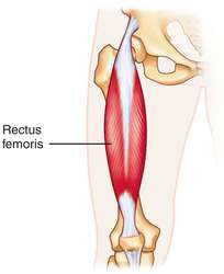
The rectus femoris muscle is the only quadriceps muscle that travels both the hip and knee. The muscle arises from the pelvis at the ASIS and AIIS and enters the other quadriceps muscles at the quadriceps tendon of the knee.

Rectus Femoris Muscle


Rectus Femoris Muscle Anatomy:

The Rectus femoris muscle is a fusiform shape & is formed in the quadriceps muscle. It is a proportion of muscle situated in the superior, anterior middle case of the thigh & is the only muscle in the quadriceps set that crosses the hip.

It is superior & overlying of the vastus intermedius muscle & superior-medial part of Vastus lateralis & Vastus medialis.

Thus the rectus femoris is its name because it runs directly down the thigh.

It is a 2-way acting muscle as it travels over the hip & knee joints; therefore, it functions to raise the knee & also helps iliopsoas in hip flexion.

A short rectus femoris may give to a higher-positioned patella about the contralateral side. A markedly compressed rectus femoris muscle is indicated by knee flexion of less than 80°or by clear dominance of the superior patellar groove.

Origin

Rectus femoris muscle originating from the anterior inferior iliac spine(AIIS) & the part of the alar of ilium superior to the acetabulum.

It has a proximal tendinous complex which is characterized by a direct tendon, an indirect tendon, & an irregular third head. Direct & indirect tendons eventually meet into a common tendon.

There is a membrane that connects the CT to the anterior superior iliac spine.

Insertion

Rectus Femoris muscle jointly with vastus medialis, vastus lateralis & vastus intermedius joins the quadriceps tendon to insert at the patella & tibial tuberosity via the patellar ligament.

Structure

It arises from two tendons: one, the anterior or straight, from the anterior inferior iliac spine; and the other, the posterior or recollected, from a groove beyond the rim of the acetabulum.

The 2 combine at an acute angle & spread into an aponeurosis that is stretched downward on the anterior surface of the muscle, & from this, the muscular fibers appear.

The muscle ends in a wide & thick aponeurosis that populates the lower 2/3rd of its posterior surface, &, slowly becoming checked into a flattened tendon, is inserted into the floor of the patella.

Function

The rectus femoris muscle, iliopsoas, and sartorius muscle are the flexors of the thigh at the hip. The rectus femoris muscle is a more vulnerable hip flexor when the knee is raised because it is already shortened & thus suffers from an active drought; the action will draft more iliacus muscle, psoas major muscle, tensor fasciae latae, and the remaining hip flexors than it will the rectus femoris.

Also, the rectus femoris muscle is not predominant in knee attachment when the hip is flexed since it is already shortened & thus suffers from an active deficit. In essence: the function of extending the knee from a seated position is primarily caused by the vastus lateralis, vastus medialis,& vastus intermedius, & less by the rectus femoris.

On the other extreme, the muscle’s capability to flex the hip & spread the knee can be compromised in a place of full hip extension & knee flexion, due to passive deficiency.

The rectus femoris muscle is a direct opponent to the hamstrings muscle, at the hip & the knee.

Relations

The proximal part of the rectus femoris muscle is found deep in the tensor fasciae latae, sartorius & iliacus muscles. All the ranges of the anterior compartment of the thigh are discovered deep in the rectus femoris. These include the capsule of the hip joint, vastus intermedius, anterior margins of vastus lateralis & vastus intermedius, lateral circumflex femoral route & some components of the femoral nerve.

Nerve supply

The neurons for volunteer thigh contraction arise near the discussion of the medial side of the precentral gyrus (the primary motor part of the brain).

These neurons send a nerve movement that is maintained by the corticospinal tract down the brainstem & spinal cord. The signal is created with the upper motor neurons carrying the signal from the precentral gyrus down through the interior capsule, via the cerebral peduncle, & into the medulla.

The nerve signal will continue down the lateral corticospinal tract until it contacts spinal nerve L4. At this part, the nerve motion will synapse from the upper motor neurons to the lower motor neurons. The signal will travel via the anterior root of L4 & into the anterior rami of the L4 nerve, leaving the spinal cord via the lumbar plexus. The posterior extension of the L4 root is the femoral nerve. 

The femoral nerve is provided by the quadriceps femoris, a 4th of which is the rectus femoris. When the rectus femoris accepts the signal that has crossed from the medial side of the precentral gyrus, it employs, expanding the knee & flexing the thigh at the hip.

Blood supply

The rectus femoris muscle is provided by the artery of the quadriceps, which can stem from 3 sources; femoral, deep femoral & lateral circumflex femoral arteries. The lateral circumflex femoral & superficial circumflex iliac routes also contribute to the blood supply of rectus femoris, but to a smaller extent.

Examination

Palpation

Rectus femoris muscle can be palpated as it is the most excellent of the quadriceps muscles. Initial palpation at the anterior inferior iliac spine, rectus femoris muscle can be handled until its insertion into the quadriceps tendon. Requesting the person to isometrically tighten the quadriceps muscle will help to determine the muscle belly.

Strength

To evaluate muscle strength for the Rectus Femoris including the rest of the quadriceps group muscle place the patient is sitting with the hip & knee flexed to 90° for grades 5, 4 & 3 while grade 2, is considered in side-lying with test limb uppermost & the knee bent to 90° position.

Wednesday, 21 June 2017

Rectus Abdominis Muscle Detail: 8 Pack Muscle

Rectus Abdominis muscle Anatomy


Rectus Abdominis Muscle
 
The rectus abdominis muscle, also known as the  "abs", this is a paired muscle running vertically on each side of the anterior wall of the human abdomen. There are two parallel muscles, separated by a midline band of connective tissue called the linea alba.

It extends from the pubic symphysis, pubic crest and pubic tubercle inferiorly, to the xiphoid process and costal cartilages of ribs V to VII superiorly. The proximal attachments are the pubic crest and the pubic symphysis. It attaches distally at the costal cartilages of ribs 5-7 and the xiphoid process of the sternum.

The rectus abdominis muscle is contained in the rectus sheath, which consists of the aponeuroses of the lateral abdominal muscles. Bands of connective tissue called the tendinous intersections traverse the rectus abdominus, which separates this parallel muscle into distinct muscle bellies.

The outer, most lateral line, defining the "abs" is the linea semilunaris. In the abdomens of people with low body fat, these bellies can be viewed externally and are commonly referred to as "four", "six", "eight", or even "ten packs", depending on how many are visible; although six is the most common.

Origin/Insertion:


The rectus abdominis is a long flat muscle, which extends along the whole length of the front of the abdomen, and is separated from its fellow of the opposite side by the linea alba.

The upper portion, attached principally to the cartilage of the fifth rib, usually has some fibers of insertion into the anterior extremity of the rib itself.

It's typically around 10 mm thick or 20 mm thick in young athletes such as handball players.

Nerve Supply

The muscles are innervated by thoraco-abdominal nerves, these are continuations of the T7-T11 intercostal nerves and p
ierce the anterior layer of the rectus sheath. Sensory supply is from the 7-12 thoracic nerves.

Blood Supply:

Several arterial blood sources supply the rectus abdominis. Categorization of the muscular vascular structure: The inferior epigastric artery and vein (or veins) supply blood to the lower portion of the muscle by running superiorly on the posterior surface of the rectus abdominis, entering the rectus fascia at the arcuate line.

Second, blood for the upper part is supplied by the superior epigastric artery, a terminal branch of the internal thoracic artery.

Lastly, the lower six intercostal arteries also contribute numerous small segmental amounts.


8 Pack Of Rectus Abdominis Muscle


Action:

The rectus abdominis is an important postural muscle. It is responsible for flexing the lumbar spine, as when doing a so-called  sit up Exercise. The rib cage is brought up to where the pelvis is when the pelvis is fixed, or the pelvis can be brought towards the rib cage (posterior pelvic tilt) when the rib cage is fixed, such as in a leg-hip raise. The two can also be brought together simultaneously when neither is fixed in space.

Sit Up Exercise is Most Common Form Of Exercise Where Rectus Abdominal Musct is Chief Muscle Used In This Action.


Six Pack Abs Workout Video:


Eight Pack Or Six Pack Exercise is most Common And Famous Among People.

Exercise Of Rectus Abdominals:

Following are the Best Exercises of Rectus Abdominis Muscle.

Sit Up Exercise:
Sit Up Exercise
Sit Up Exercise

In Supine Position Flexes Both Legas , Hands Behind Head And Flexes The Spine And Heads Towards Knee And Repetation Of Same.
Sit Up Exercise
Sit Up Exercise 


Bilateral Leg Elevation In Supine Postion Uses Lower Abdominals Muscle.
Rectus Abdominis is strong Back Flexor Muscle.


Knee To Chest Exercise


To Reduce Lordosis Strengthening Of Rectus Abdominis Muscle is required.
strengthening of Rectus Abdominis And Other Back Flexor Muscle in Back Pain is Most Common And is Called Williams Abdominal Exercise.

Anatomical Variations

A variation of the rectus abdominis or pectoralis major may be the sternalis muscle. Occasionally, some fibers are joined to the side of the xiphoid process and the costoxiphoid ligaments.

Importance of the Muscles

The abdominalis recti, a muscle of the core, contribute to core stability. The rectus abdominis, transversus abdominis, erector spinae, and obliques are among the core muscles that work in collaboration to prevent lower back injuries. They look like a natural weight belt. People who have weak core muscles are more likely to have spinal issues.

At the linea alba, there is a midline separation known as diastasis recti abdominis. Diastasis is defined as any visible bulging during physical exertion or a palpable midline gap greater than 2.5 cm. It typically develops around the umbilicus, but it can occur anywhere between the pubic bone and the xiphoid process.

This spread belly is also common in newborns, and it should go away on its own. The majority of premature and African American infants exhibit it.

Assessment

Palpation

To ensure that the patient's muscles are relaxed, place a pillow under their knees and place them in a supine position. Palpate the patient's muscles along their xiphoid process, adjacent ribs, and pubis bone on both sides. After encouraging the patient to lift their trunk, ask them to relax.

Muscle Power Testing

It is more important to evaluate the strength of the abdominal muscles than their flexibility because weak abdominal muscles can lead to serious issues down the road.

When performing MMT for rectus abdominis, the patient is typically asked to raise his or her trunk against gravity while supine until the inferior angles of the scapula are off the table. The therapist then begins to grade the patient based on the patient's performance and capacity to complete the exercise as directed.

Clinical Significance

An injury to one of the abdominal wall's muscles is known as an abdominal muscle strain, sometimes known as a pulled abdominal muscle. Excessive stretching of the muscle results in a strain. The muscle fibers tear when this happens. Usually, a strain results in tiny tears within the muscle, but in extreme cases, the muscle may separate from its attachment.

An accumulation of blood in the rectus abdominis muscle's sheath is known as a rectus sheath hematoma. It can cause pain in the abdomen with or without a mass. Either a muscle tear or an epigastric artery rupture could be the source of the hematoma. Anticoagulation, coughing, pregnancy, abdominal surgery, and trauma are some of the causes of this.

There is evidence that the once-benign condition is becoming more common and serious due to the aging population and the widespread use of anticoagulant medications.

Individuals may have a positive Carnett's sign upon abdominal examination.

Although they usually go away on their own without treatment, hematomas can take months to go away.