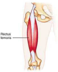
The rectus femoris muscle is the only quadriceps muscle that travels both the hip and knee. The muscle arises from the pelvis at the ASIS and AIIS and enters the other quadriceps muscles at the quadriceps tendon of the knee.
 |
| Rectus Femoris Muscle |
Rectus Femoris Muscle Anatomy:
The Rectus femoris muscle is a fusiform shape & is formed in the quadriceps muscle. It is a proportion of muscle situated in the superior, anterior middle case of the thigh & is the only muscle in the quadriceps set that crosses the hip.
It is superior & overlying of the vastus intermedius muscle & superior-medial part of Vastus lateralis & Vastus medialis.
Thus the rectus femoris is its name because it runs directly down the thigh.
It is a 2-way acting muscle as it travels over the hip & knee joints; therefore, it functions to raise the knee & also helps iliopsoas in hip flexion.
A short rectus femoris may give to a higher-positioned patella about the contralateral side. A markedly compressed rectus femoris muscle is indicated by knee flexion of less than 80°or by clear dominance of the superior patellar groove.
Origin
Rectus femoris muscle originating from the anterior inferior iliac spine(AIIS) & the part of the alar of ilium superior to the acetabulum.
It has a proximal tendinous complex which is characterized by a direct tendon, an indirect tendon, & an irregular third head. Direct & indirect tendons eventually meet into a common tendon.
There is a membrane that connects the CT to the anterior superior iliac spine.
Insertion
Rectus Femoris muscle jointly with vastus medialis, vastus lateralis & vastus intermedius joins the quadriceps tendon to insert at the patella & tibial tuberosity via the patellar ligament.
Structure
It arises from two tendons: one, the anterior or straight, from the anterior inferior iliac spine; and the other, the posterior or recollected, from a groove beyond the rim of the acetabulum.
The 2 combine at an acute angle & spread into an aponeurosis that is stretched downward on the anterior surface of the muscle, & from this, the muscular fibers appear.
The muscle ends in a wide & thick aponeurosis that populates the lower 2/3rd of its posterior surface, &, slowly becoming checked into a flattened tendon, is inserted into the floor of the patella.
Function
The rectus femoris muscle, iliopsoas, and sartorius muscle are the flexors of the thigh at the hip. The rectus femoris muscle is a more vulnerable hip flexor when the knee is raised because it is already shortened & thus suffers from an active drought; the action will draft more iliacus muscle, psoas major muscle, tensor fasciae latae, and the remaining hip flexors than it will the rectus femoris.
Also, the rectus femoris muscle is not predominant in knee attachment when the hip is flexed since it is already shortened & thus suffers from an active deficit. In essence: the function of extending the knee from a seated position is primarily caused by the vastus lateralis, vastus medialis,& vastus intermedius, & less by the rectus femoris.
On the other extreme, the muscle’s capability to flex the hip & spread the knee can be compromised in a place of full hip extension & knee flexion, due to passive deficiency.
The rectus femoris muscle is a direct opponent to the hamstrings muscle, at the hip & the knee.
Relations
The proximal part of the rectus femoris muscle is found deep in the tensor fasciae latae, sartorius & iliacus muscles. All the ranges of the anterior compartment of the thigh are discovered deep in the rectus femoris. These include the capsule of the hip joint, vastus intermedius, anterior margins of vastus lateralis & vastus intermedius, lateral circumflex femoral route & some components of the femoral nerve.
Nerve supply
The neurons for volunteer thigh contraction arise near the discussion of the medial side of the precentral gyrus (the primary motor part of the brain).
These neurons send a nerve movement that is maintained by the corticospinal tract down the brainstem & spinal cord. The signal is created with the upper motor neurons carrying the signal from the precentral gyrus down through the interior capsule, via the cerebral peduncle, & into the medulla.
The nerve signal will continue down the lateral corticospinal tract until it contacts spinal nerve L4. At this part, the nerve motion will synapse from the upper motor neurons to the lower motor neurons. The signal will travel via the anterior root of L4 & into the anterior rami of the L4 nerve, leaving the spinal cord via the lumbar plexus. The posterior extension of the L4 root is the femoral nerve.
The femoral nerve is provided by the quadriceps femoris, a 4th of which is the rectus femoris. When the rectus femoris accepts the signal that has crossed from the medial side of the precentral gyrus, it employs, expanding the knee & flexing the thigh at the hip.
Blood supply
The rectus femoris muscle is provided by the artery of the quadriceps, which can stem from 3 sources; femoral, deep femoral & lateral circumflex femoral arteries. The lateral circumflex femoral & superficial circumflex iliac routes also contribute to the blood supply of rectus femoris, but to a smaller extent.
Examination
Palpation
Rectus femoris muscle can be palpated as it is the most excellent of the quadriceps muscles. Initial palpation at the anterior inferior iliac spine, rectus femoris muscle can be handled until its insertion into the quadriceps tendon. Requesting the person to isometrically tighten the quadriceps muscle will help to determine the muscle belly.
Strength
To evaluate muscle strength for the Rectus Femoris including the rest of the quadriceps group muscle place the patient is sitting with the hip & knee flexed to 90° for grades 5, 4 & 3 while grade 2, is considered in side-lying with test limb uppermost & the knee bent to 90° position.
No comments:
Post a Comment