Sunday, 28 January 2018

Bicipital Tendinitis: And Physiotherapy Exercise

Biceps tendinitis
Bicipital Tendinitis 

Biceps tendinitis is inflammation of the tendon around the long head of the biceps muscle.

Biceps tendinitis is a common injury that occurs when the muscles and connective tissues of the arm become inflamed and swollen due to overuse. The biceps muscle is located in the upper arm, and it helps with both flexion and rotation of the shoulder joint. The tendons that attach the biceps muscle to the bones of the shoulder are also susceptible to injury, which can lead to biceps tendinitis.

Overview:

Biceps tendinosis is caused by degeneration of the tendon from athletics requiring overhead motion or from the normal aging process.

Inflam-mation of the biceps tendon in the bicipital groove, which is known as primary biceps tendinitis, occurs in 5 percent of patients with biceps tendinitis.

Biceps tendinitis and tendinosis are commonly accompanied by rotator cuff tears or SLAP (superior labrum anterior to posterior) lesions. Patients with biceps tendinitis or tendinosis usually complain of a deep, throbbing ache in the anterior shoulder. Repetitive overhead motion of the arm initiates or exacerbates the symptoms. 

The most common isolated clinical finding in biceps tendinitis is bicipital groove point tenderness with the arm in 10 degrees of internal rotation. Local anesthetic injections into the biceps tendon sheath may be therapeutic and diagnostic. Ultrasonography is preferred for visualizing the overall tendon, whereas magnetic resonance imaging or computed tomography arthrography is preferred for visualizing the intraarticular tendon and related pathology. 

Conservative management of biceps tendinitis consists of rest, ice, oral analgesics, physical therapy, or corticosteroid injections into the biceps tendon sheath. Surgery should be considered if conservative measures fail after three months, or if there is severe damage to the biceps tendon.

Anatomy and Physiology:

The long head of the biceps tendon rises from the supraglenoid tubercle and the superior glenoid labrum

The proximal portion of the long head of the biceps tendon is extrasynovial but intra-articular.
5 The tendon travels obliquely inside the shoulder joint, across the humeral head anteriorly, and exits the joint within the bicipital groove of the humeral head beneath the transverse humeral ligament
.
 
The bicipital groove is defined by the greater tuberosity (lateral) and the lesser tuberosity (medial). The biceps tendon is contained in the rotator interval, a triangular area between the subscapularis and supraspinatus tendons at the shoulder (Figure 1). The rotator interval is responsible for keeping the biceps tendon in its correct location.6–8 Because the rotator interval is usually indistinguishable from the rotator cuff and capsule, lesions of the biceps tendon are usually accompanied by lesions of the rotator cuff.

SLAP lesions are often present in patients with biceps tendinitis and tendinosis. The anterosuperior labrum and superior labrum are more likely to tear than the inferior portion of the labrum because they are not attached as tightly to the glenoid.9–13 Additionally, certain conditions that affect the glenohumeral joint may also involve the biceps tendon because it is intra-articular. These may include rheumatologic (e.g., rheumatoid arthritis, lupus), infectious, or other types of reactive or inflammatory conditions.

Symptoms:
Bicipital Tendinitis 

Patients with biceps tendinitis often complain of a deep, throbbing ache in the anterior shoulder. The pain is usually localized to the bicipital groove and may radiate toward the insertion of the deltoid muscle, or down to the hand in a radial distribution.

This makes it difficult to distinguish from pain that is secondary to impingement or tendinitis of the rotator cuff, or cervical disk disease. Pain from biceps tendinitis usually worsens at night, especially if the patient sleeps on the affected shoulder.

Repetitive overhead arm motion, pulling, or lifting may also initiate or exacerbate the pain.9 The pain is most noticeable in the follow-through of a throwing motion.3 Instability of the tendon may present as a palpable or audible snap when range of motion of the arm is tested.

Rupture of the biceps tendon is one of the most common musculotendinous tears. If the biceps has ruptured, patients will describe an audible, painful popping, followed by relief of symptoms. The anterior shoulder may be bruised, with a bulge visible above the elbow as the muscle retracts distally from the rupture point. Risk factors of biceps rupture include a history of rotator cuff tear, recurrent tendinitis, contralateral biceps tendon rupture, rheumatoid arthritis, age older than 40 years, and poor conditioning.9 If a patient has a feeling of popping, catching, or locking in the shoulder, a SLAP lesion may be present. This usually occurs after trauma, such as a direct blow to the shoulder, a fall on an outstretched arm, or repetitive overhead motion in athletes.

The most common finding of biceps tendon injury is bicipital groove point tenderness.


PHYSICAL EXAMINATION:

Many provocative tests (i.e., Yergason, Neer, Hawkins, and Speed tests) have been developed to isolate pathology of the biceps tendonhowever, because these tests create impingement underneath the coracoacromial arch, it is difficult to rule out concomitant rotator cuff lesions.

The Yergason test requires the patient to place the arm at his or her side with the elbow flexed at 90 degrees, and supinate against resistance18 (Figure 2). The test is considered positive if pain is referred to the bicipital groove.

The Neer test involves internal rotation of the arm while in the forward flexed position16. If the patient experiences pain, it is a positive sign of impingement syndrome.

During the Hawkins test, the patient flexes the elbow to 90 degrees while the physician elevates the patient's shoulder to 90 degrees and places the forearm in a neutral position19 (Figure 4). With the arm supported, the humerus is rotated internally. The test is positive if bicipital groove pain is present.


Speed test, the patient tries to flex the shoulder against resistance with the elbow extended and the forearm supinated9,20 (Figure 5). A positive test is pain radiating to the bicipital groove. If any of these tests is positive, it indicates that impingement is present, which can lead to biceps tendinitis or tendinosis.

Advantages and Disadvantages of Radiologic Imaging Studies in the Evaluation of Biceps Tendinitis.

IMAGING STUDY : 

Arthrography (used with MRI or CT to visualize the joint capsule and glenoid labrum)

ADVANTAGES

CT arthrography shows biceps tendon subluxations, ruptures, dislocations, and SLAP lesion
MRI arthrography is preferable for diagnosing biceps lesions and SLAP lesions14 because the agreement between MRI and arthroscopy for biceps lesions is only 37 percent and 60 percent for rotator cuff lesions

DISADVANTAGES

Invasive
Filling of the biceps tendon sheath is unreliable
Sharp images of the tendon may be lost


Ionizing radiation
Bicipital groove view radiography

ADVANTAGES

Shows the width and medial wall angle of the bicipital groove, spurs in the groove, and supertubercular bone spur or ridge
Inexpensive


DISADVANTAGES

Does not show possible intra-articular disorders of the labrum (soft tissue injuries)

MRI

ADVANTAGES

Excellent evaluation of the superior labral complex and biceps tendon

DISADVANTAGES

Partial tears of the biceps tendon are more difficult to detect than complete ruptures
Expensive
Poorly
 

Treatment :


CONSERVATIVE:

Biceps tendinitis or tendinosis may respond to analgesia with nonsteroidal anti-inflammatory drugs (NSAIDs).

Physiotherapy Treatment:


Bicipital Tendinitis And Exercise


Ice, rest from overhead activity, or physical therapy. Rehabilitation of an athlete's shoulder involves four phases:

Rest; stretching exercises of the scapula, rotator cuff, and posterior capsule; 

The goal of stretching is to regain a balanced range of motion without stiffness or pain in any position.

Taping Over Biceps Give Great Relief From Pain And Allow Smooth Movement.


Tapping in Bicipital Tendinitis


Strengthening and a progressively difficult throwing program.
The patient may begin exercises after the shoulder is pain-free.
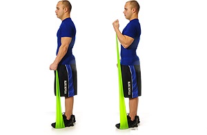


Strengthening Exercise Of Biceps Muscle


Monday, 22 January 2018

DEEP VEIN THROMBOSIS : OVERVIEW & TREATMENT

Profound vein thrombosis (DVT) happens when a blood coagulation (thrombus) frames in at least one of the profound veins in your body,
DVT  
as a rule in your legs. Profound vein thrombosis can cause leg torment or swelling, yet in addition can happen without any side effects. Profound vein thrombosis can be intense on the grounds that blood coagulations in your veins can loosen up, go through your circulatory system and cabin in your lungs, blocking blood stream (pneumonic embolism). 

PATHOPHYSIOLOGY:
Deep Vein Thrombosis 


Lower-appendage profound venous thrombosis (DVT) influences between 1% to 2% of hospitalized patients. These thrombi upset the vascular uprightness of the lower appendages and are the wellspring of emboli that

slaughter around 200,000 patients every year in the United States. The reasons for thrombosis incorporate vessel divider harm, stasis or low stream, and hypercoagulability. These components support cluster arrangement by disturbing the adjust of the contradicting coagulative and fibrinolytic frameworks.

Indications

Profound vein thrombosis signs and indications can include:

Swelling in the influenced leg. Once in a while, there's swelling in the two legs.

Torment in your leg. The torment regularly begins in your calf and can have a craving for cramping or soreness.

Red or stained skin on the leg.

A sentiment warmth in the influenced leg.

Profound vein thrombosis can happen without recognizable side effects.

The notice signs and side effects of an aspiratory embolism include:

Sudden shortness of breath

Chest agony or distress that compounds when you take a full breath or when you hack

Feeling woozy or mixed up, or blacking out

Fast heartbeat

Hacking up blood

Causes

The blood coagulations of profound vein thrombosis can be caused by anything that keeps your blood from coursing or thickening ordinarily, for example, damage to a vein, surgery, certain solutions and restricted development.

Hazard factors

Numerous elements can build your danger of growing profound vein thrombosis (DVT). The more you have, the more prominent your danger of DVT. Hazard factors include:

Acquiring a blood-thickening issue. A few people acquire a turmoil that influences their blood to cluster all the more effortlessly. This condition without anyone else won't not cause blood clusters unless joined with at least one other hazard factors.

Drawn out bed rest, for example, amid a long clinic stay, or loss of motion. At the point when your legs stay still for long stretches, your lower leg muscles don't contract to enable blood to course, which can build the danger of blood clusters.

Damage or surgery. Damage to your veins or surgery can build the danger of blood clusters.

Pregnancy. Pregnancy builds the weight in the veins in your pelvis and legs. Ladies with an acquired thickening issue are particularly in danger. The danger of blood clumps from pregnancy can proceed for up to a month and a half after you have your child.

Anti-conception medication pills (oral contraceptives) or hormone substitution treatment. Both can expand your blood's capacity to clump.

Being overweight or fat. Being overweight expands the weight in the veins in your pelvis and legs.

Smoking. Smoking influences blood thickening and flow, which can build your danger of DVT.

Growth. A few types of disease increment substances in your blood that reason your blood to clump. A few types of disease treatment additionally increment the danger of blood clusters.

Heart disappointment. This expands your danger of DVT and pneumonic embolism. Since individuals with heart disappointment have restricted heart and lung work, the side effects caused by even a little aspiratory embolism are more observable.

Incendiary inside illness. Gut ailments, for example, Crohn's sickness or ulcerative colitis, increment the danger of DVT.

An individual or family history of profound vein thrombosis or pneumonic embolism. In the event that you or somebody in your family has had either of these, you may be at more serious danger of building up a DVT.

Age. Being more established than 60 expands your danger of DVT, however it can happen at any age.

Sitting for drawn out stretches of time, for example, when driving or flying. At the point when your legs stay still for quite a long time, your lower leg muscles don't contract, which regularly enables blood to circle. Blood clumps can shape in the calves of your legs if your lower leg muscles don't move for long stretches.

Confusions

Pneumonic embolism

Pneumonic embolism

A genuine confusion related with profound vein thrombosis is aspiratory embolism.

Pneumonic embolism

A pneumonic embolism happens when a vein in your lung ends up noticeably hindered by a blood coagulation (thrombus) that movements to your lung from another piece of your body, for the most part your leg.

An aspiratory embolism can be dangerous. It's vital to look for signs and manifestations of a pneumonic embolism and look for medicinal consideration in the event that they happen. Signs and side effects of a pneumonic embolism include:

Sudden shortness of breath

Chest torment or distress that intensifies when you take a full breath or when you hack

Feeling bleary eyed or unsteady, or swooning

Fast heartbeat

Hacking up blood

Postphlebitic disorder

A typical complexity that can happen after profound vein thrombosis is known as postphlebitic disorder, additionally called postthrombotic disorder. Harm to your veins from the blood coagulation diminishes blood stream in the influenced regions, which can cause:

Determined swelling of your legs (edema)

Leg torment

Skin staining

Skin wounds

Avoidance

Measures to avoid profound vein thrombosis include:

Abstain from sitting still. In the event that you have had surgery or have been on bed rest for different reasons, endeavor to move at the earliest opportunity. In case you're sitting for some time, don't fold your legs, which can hamper blood stream. In case you're voyaging a long separation via auto, stop each hour or thereabouts and stroll around.

In case you're on a plane, stand or walk every so often. In the event that you can't do that, activity your lower legs. Take a stab at raising and bringing down your foot rear areas while keeping your toes on the floor, at that point raising your toes with your rear areas are on the floor.

Roll out way of life improvements. Get more fit and quit smoking.

Exercise. Customary exercise brings down your danger of blood clumps, which is particularly essential for individuals who sit a ton or travel much of the time.

Conclusion

To analyze profound vein thrombosis, your specialist will get some information about your side effects. You'll additionally have a physical exam so your specialist can check for regions of swelling, delicacy or staining on your skin. Contingent upon the fact that you are so prone to have a blood coagulation, your specialist may recommend tests, including:

Ultrasound. A wandlike gadget (transducer) set over the piece of your body where there's a coagulation sends sound waves into the region. As the sound waves go through your tissue and reflect back, a PC changes the waves into a moving picture on a video screen. A coagulation may be noticeable in the picture.

Some of the time a progression of ultrasounds are done more than a few days to decide if a blood coagulation is developing or to check for another one.

Blood test. All individuals who create serious profound vein thrombosis have a hoisted blood level of a substance called D dimer.

Venography. A color is infused into an expansive vein in your foot or lower leg. A X-beam makes a picture of the veins in your legs and feet, to search for clusters. Be that as it may, less intrusive strategies for finding, for example, ultrasound, can as a rule affirm the determination.

CT or MRI checks. Either can give visual pictures of your veins and may appear in the event that you have a coagulation. Here and there these sweeps performed for different reasons uncover a coagulation.

Treatment : 

Bolster leggings

Stockings in DVT


Bolster tights

Profound vein thrombosis (DVT) treatment is gone for keeping the coagulation from getting greater and keeping it from loosening up and causing an aspiratory embolism. At that point the objective progresses toward becoming lessening your odds of profound vein thrombosis happening once more.

Profound vein thrombosis treatment alternatives include:

Blood thinners. Profound vein thrombosis is most regularly treated with anticoagulants, likewise called blood thinners. These medications, which can be infused or taken as pills, diminish your blood's capacity to cluster. They don't separate existing blood clumps, however they can keep clusters from getting greater and decrease your danger of growing more clumps.

The injectable prescriptions can be given as a shot under the skin or by infusion into your arm vein (intravenous).

Heparin is regularly given intravenously. Other comparative blood thinners, for example, enoxaparin (Lovenox), dalteparin (Fragmin) or fondaparinux (Arixtra), are infused under the skin.

You may get an injectable blood more slender for a couple of days, after which pills, for example, warfarin (Coumadin, Jantoven) or dabigatran (Pradaxa) are begun. When warfarin has diminished your blood, the injectable blood thinners are ceased.

Other blood thinners can be given in pill shape without the requirement for an injectable blood more slender. These incorporate rivaroxaban (Xarelto), apixaban (Eliquis) or edoxaban (Savaysa).

You may need to take blood more slender pills for three months or more. It's essential to take them precisely as your specialist educates in light of the fact that taking excessively or too little can cause genuine reactions.

On the off chance that you take warfarin, you'll require occasional blood tests to check to what extent it takes your blood to cluster. Pregnant ladies shouldn't take certain blood-diminishing meds.

Cluster busters. On the off chance that you have a more genuine kind of profound vein thrombosis or aspiratory embolism, or if different meds aren't working, your specialist may endorse drugs that separate coagulations immediately, called cluster busters or thrombolytics.

These medications are either given through an IV line to separate blood clumps or through a catheter put straightforwardly into the coagulation. These medications can cause genuine dying, so they're for the most part held for extreme instances of blood clumps.

Channels. In the event that you can't take drugs to thin your blood, you may have a channel embedded into an extensive vein — the vena cava — in your guts. A vena cava channel keeps clusters that loosen up from hotel in your lungs.

Pressure leggings. To assist counteract swelling related with profound vein thrombosis, these are worn on your legs from your feet to about the level of your knees.

Monday, 1 January 2018

Torticollis And Physiotherapy Treatment Overview :

TORTICOLIS 
Torticollis 


Torticolis is a condition ( Also Called ‘wryneck’) in which the baby’s head is tilted One Side. The head often rotates towards one shoulder and tilts away to the opposite side. The term ‘congenital’ is also sometime used when describing From Birth torticollis. This means that it is present at or shortly after birth. Babies treated early with physiotherapy programmes usually Give Good Result to treatment.

Torticollis is a symptom related to turning or bending of the neck one side . Many different causes are possible. In newborns, torticollis usually results from injury during labor and delivery or the infant’s position in the womb. Less often, it is caused by birth defects. In older children, torticollis may result from injuries to the neck muscles, common infections, or other causes.
Painful spasms of the neck muscles may occur.
Other symptoms may be present, depending on the cause. For example, there may be a tender lymph node (gland) if the cause is infection.


TYPES:
  • Congenital torticolis.
  • Acquired torticolis.
Acquired torticolis


ANATOMY:

The normal physiologic range of rotation of the atlas on the axis is 25-53 degrees to either side. The transverse ligament is the primary stabilizer of the atlantoaxial joint and prevents excessive anterior motion of the atlas on the axis. It extends behind the dens, between the medial portions of the lateral masses of C1. The paired alar ligaments act as secondary stabilizers to prevent anterior shift. The alar ligaments extend from the lateral aspect of the dens tip to the medial aspect of the occipital condyles, with a lower portion attaching to the medial aspect of the lateral masses of C1.

The sternocleidomastoid muscle has a sternal and clavicular head. The sternal head is directed from the manubrium sterni superiorly, laterally and posteriorly and the clavicular from the medial third of the clavicle vertically upward. It runs to the mastoid process. It enables an ipsilateral lateral flexion and a contralateral rotation. The muscle extends the upper part of the cervical spine and flexes the lower part.


ETIOLOGY:

Muscular in more than 80% of the cases. Types muscular torticollis
          - Fibromatosis colli: torticollis with palpable mass in the SCM;
          - Tightness of the SCM without an apparent mass;
Postural torticollis with neither mass or tightness.
Birth trauma: facet dislocation, tears in the sternocleidomastoid muscle
Congenital anomalies of the craniovertebral junction: occipitoatlantal fusion or Klippel-Feil syndrome.
Sternocleidomastoid tumour.
Ocular abnormalities.
Intrauterine mechanical factors


CAUSES:


Congenital Torticollis 


Sitting or sleeping in an unusual position without adequate neck support.
Poor posture when looking at a computer screen.
Carrying heavy unbalanced loads (for example, a briefcase or shopping bag).
Allowing certain muscles of the neck to be exposed to cold (sleeping in a draught).


SYMPTOMS:


SternocleidoMustoid Muscle 


The twisting of your neck (torticollis) occurs when your muscles supporting the neck on one side are painful.
The pain is usually on one side of your neck and stiffness of the muscles in that area twists the neck to one side. You may find it very difficult when you try to straighten your neck, due to pain. Occasionally, the pain is in the middle of your neck.

The pain may spread to the back of your head or to your shoulder. The muscles of your affected side may be tender. Pressure on certain areas may trigger a 'spasm' of these muscles. Movement of your neck is restricted, particularly on one side.


DIAGNOSIS:

A thorough neurologic examination should be performed, and anteroposterior and lateral radiographs of the cervical spine should be obtained. A CT scan or MRI of the head and neck is necessary for any patient with persistent neck pain or with neurologic signs and symptoms.

TREATMENTS:

Medicines:
Pain killer are often helpful. such as,
Paracetamol at strength is often sufficient.
Anti inflammatory painkillers.
A stronger pain killer such codeine.
A muscle relaxant such as diazepam.

Other treatments such as:
Rest.
A good posture.
A firm supporting pillow.
Heat pack.


PHYSIOTHERAPY TREATMENTS AND EXERCISES:


Exercise In Sternocleidomustoid Muscle


  • Positioning.
  • Gentle range of motion exercises for neck.
  • Stretching of sternocleido mastoid muscle.
  • Strengthening exercises.
  • Activities to encourage active head movement.
  • Visual tracking.
  • Lateral head tilt.
  • Therapy ball exercises.
  • Side sitting exercises.
  • Hands and knees.
  • Kneeling to standing.
  • Assisted rolling.
  • Proper sidelying.

Torticollis treatment at home for babies:

The best method of torticollis treatment is to encourage your baby to turn his or her head in both directions. This will help to loosen tense neck muscles and tighten the loose ones. Here are some exercises to try:
When your baby wants to eat, offer the bottle or your breast in a way that encourages your baby to turn away from the favored side. (Use your child's desire to eat to encourage him or her along!)
When putting your baby down to sleep, position him or her to face the wall. Since babies prefer to look out onto the room, your baby will actively turn away from the wall and this will stretch the tightened muscles of the neck. 
During play, draw your baby's attention with toys and sounds to make him or her turn in both directions.


Other Deformity Related Article:

CTEV Detail

 
Radial Nerve Palsy
 Radial Nerve Palsy