Friday, 12 September 2014

RHEUMATOID ARTHRITIS : PHYSIOTHERAPY Treatment :

RHEUMATOID ARTHRITIS AND ITS PHYSIOTHERAPY TREATMENT.:
Rheumatoid Arthritis

Rheumatoid arthritis (RA) is an autoimmune disease that results in a chronic, systemic inflammatory disorder that may affect many tissues and organs, but principally attacks flexible (synovial) joints. It can be a disabling and painful condition, which can lead to substantial loss of functioning and mobility if not adequately treated

Rheumatoid arthritis : 


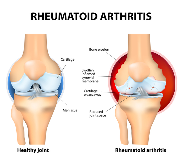
Rheumatoid Arthritis Joint


Rheumatoid arthritis is a disorder in which the body's own immune system starts to attack body tissues. The attack is not only directed at the joint but to many other parts of the body. In rheumatoid arthritis, most damage occurs to the joint lining and cartilage which eventually results in erosion of two opposing bones. Rheumatoid arthritis often affects joints in the fingers, wrists, knees and elbows. The disease is symmetrical (appears on both sides of the body) and can lead to severe deformity in a few years if not treated. Rheumatoid arthritis occurs mostly in people aged 20 and above. In children, the disorder can present with a skin rash, fever, pain, disability, and limitations in daily activities. Often, it is not clear why the rheumatoid arthritis occurred. With earlier diagnosis and aggressive treatment, many individuals can lead a decent quality of life. The drugs to treat rheumatoid arthritis range from corticosteroids to monoclonal antibodies given intravenously.In rare cases, surgery may be required to replace joints but there is no cure for the illness.


Normal Joint-OA Knee- RA Knee


signs and symptoms of rheumatoid arthritis may include:

  •     Tender, warm, swollen joints
  •     Morning stiffness that may last for hours
  •     Firm bumps of tissue under the skin on your arms (rheumatoid nodules)
  •     Fatigue, fever and weight loss
Early rheumatoid arthritis tends to affect your smaller joints first — particularly the joints that attach your fingers to your hands and your toes to your feet. As the disease progresses, symptoms often spread to the knees, ankles, elbows, hips and shoulders. In most cases, symptoms occur in the same joints on both sides of your body.
Hands In RA

Rheumatoid arthritis signs and symptoms may vary in severity and may even come and go. Periods of increased disease activity, called flares, alternate with periods of relative remission — when the swelling and pain fade or disappear. Over time, rheumatoid arthritis can cause joints to deform and shift out of place.
When to see a doctor

Make an appointment with your doctor if you have persistent discomfort and swelling in your joints.


CAUSES :


Rheumatoid arthritis occurs when your immune system attacks the synovium — the lining of the membranes that surround your joints. The resulting inflammation thickens the synovium, which can eventually destroy the cartilage and bone within the joint. The tendons and ligaments that hold the joint together weaken and stretch. Gradually, the joint loses its shape and alignment.

Doctors don't know what starts this process, although a genetic component appears likely. While your genes don't actually cause rheumatoid arthritis, they can make you more susceptible to environmental factors — such as infection with certain viruses and bacteria — that may trigger the disease.

RISK FACTORS

Factors that may increase your risk of rheumatoid arthritis include:

    Your sex. Women are more likely to develop rheumatoid arthritis than men are.
    Age. Rheumatoid arthritis can occur at any age, but it most commonly begins between the ages of 40 and 60.
    Family history. If a member of your family has rheumatoid arthritis, you may have an increa
sed risk of the disease.

   Examination Of RA( RHEUMATOID ARTHRITIS) :
 
        Blood tests for rheumatoid arthritis

          Blood tests can measure inflammation. You may have one of these tests:

    erythrocyte sedimentation rate (ESR)
    C-reactive protein (CRP).

Both of these may show a high value when inflammation is present.

Blood tests can show if you’re anaemic and may be used to detect rheumatoid factor, which is an antibody produced by a reaction in your immune system.


X-RAY :


X-rays will show any damage caused to your joints by the inflammation in rheumatoid arthritis. The changes often show up in x-rays of your feet before they appear in other joints, so your doctor may want to x-ray your feet even if they’re not causing you any problems.
-RHEUMATOID FACTOR TEST


COMPLICATIONS : 

Rheumatoid arthritis increases your risk of developing:

    Osteoporosis. Rheumatoid arthritis itself, along with some medications used for treating rheumatoid arthritis, can increase your risk of osteoporosis — a condition that weakens your bones and makes them more prone to fracture.
    Carpal tunnel syndrome. If rheumatoid arthritis affects your wrists, the inflammation can compress the nerve that serves most of your hand and fingers.
    Heart problems. Rheumatoid arthritis can increase your risk of hardened and blocked arteries, as well as inflammation of the sac that encloses your heart.
    Lung disease. People with rheumatoid arthritis have an increased risk of inflammation and scarring of the lung tissues, which can lead to progressive shortness of breath.


 PHYSIOTHERAPY TREATMENT :

Exercise In Rheumatoid Arthritis


Physiotherapy treatment is important in helping patients with RA manage their disease. In conjunction with occupational therapists, physiotherapists educate patients in joint protection strategies, use of assistive devices, and performance of therapeutic exercises.
  • Rest and Splinting. The joints should be put into rest during the acute stage of the disease. Bed rest relieves the pain in cases of extensive joint involvement


  • Compression Gloves. Patients using compression gloves have reported reduced joint swelling and increased well-being


  • Assistive Devices and Adaptive Equipment. Assistive devices are used in order to reduce functional deficits, to diminish pain, and to keep patients' independence and self-efficiency. Loading over the hip joint may be reduced by 50% by holding a cane.


  • Massage Therapy. Massage is a commonly used treatment tool that improves flexibility, enhances a feeling of connection with other treatment modalities, improves general well being, and can help to diminish swelling of inflamed joints.


  • Aerobic Exercise
Physiotherapy Exercise 


Therapeutic Exercise :


Muscle weakness in patients with RA may occur because of immobilization or reduction in activities of daily living. Maintenance of normal muscle strength is important not only for physical function but also for stabilization of the joints and prevention of traumatic injuries. It may be proposed that exercise therapy has beneficial effects on increasing physical capacity rather than reducing the activity of the disease.

  • There should be no straining exercises during the acute arthritis. However, every joint should be moved in the ROM at least once per day in order to prevent contracture. In the case of acutely inflamed joints, isometric exercises provide adequate muscle tone


  • Moderate contractures should be held for 6 seconds and repeated 5-10 times each day.


  • Low-intensity isokinetic knee exercises (by 50% of the maximum voluntary contraction) were reported to be safe and effective in patients with RA
patients with active arthritis should particularly avoid activities such as climbing stairs or weight lifting. Producing excessive stress over the tendons during the stretching exercises should be avoided. In sudden stretches, tendons or joint capsules may be damaged. Finally, in chronic stage with inactive arthritis, conditioning exercises such as swimming, walking, and cycling with adequate resting periods are recommended. They increase muscle endurance and aerobic capacity and improve functions of the patient in general, and they also make the patient feel better.

 Related Post :


Tennis Elbow And Physiotherapy


Ads :


Post Free Ads Online


Osteoarthritis and Physical Therapy Treatment , Exercise


Osteoarthritis of Knee
Arthritis is a general term that means inflammation of the joints. Osteoarthritis, commonly known as wear and tear arthritis, is the most common type of arthritis. It is associated with a breakdown of cartilage in joints and can occur in almost any joint in the body. It commonly occurs in the weight-bearing joints of the hips, knees, and spine. It also affects the fingers, thumb, neck, and large toe.
wear and tear arthritis, is the most common type of arthritis. It is associated with a breakdown of cartilage in joints and can occur in almost any joint in the body. It commonly occurs in the weight-bearing joints of the hips, knees, and spine. It also affects the fingers, thumb, neck, and large toe.

Osteoarthritis -- also called OA -- usually does not affect other joints unless previous injury , excessive stress or an underlying disorder of cartilage is involved.

Cartilage is a firm, rubbery material that covers the ends of bones in normal joints. Its main function is to reduce friction in the joints and serve as a "shock absorber." The shock-absorbing quality of normal cartilage comes from its ability to change shape when compressed (flattened or pressed together).

Osteoarthritis causes the cartilage in a joint to become stiff and lose its elasticity, making it more susceptible to damage. Over time, the cartilage may wear away in some areas, greatly decreasing its ability to act as a shock absorber. As the cartilage deteriorates, tendons and ligaments stretch, causing pain. If the condition worsens, the bones could rub against each other.

WHO GETS OSTEOARTHRITIS?


 The chance of developing the disease increases with age. Most people over age 60 have osteoarthritis to some degree, but its severity varies. Even people in their 20s and 30s can get osteoarthritis, although there is often an underlying reason, such as joint injury or repetitive joint stress from overuse. In people over age 50, more women than men have osteoarthritis.

WHAT ARE THE SYMPTOMS OF OSTEOARTHRITIS?


Symptoms of osteoarthritis most often develop gradually and include:


  • Joint aching and soreness, especially with movement
  • Pain after overuse or after long periods of inactivity
  • Joint Stiffness after periods of rest
  • Bony enlargements in the middle and end joints of the fingers (which may or may not be painful)
  • Joint swelling
  • Muscles around joint are become weak.
  • Reduced range of motion and loss of use of the joint.
  • Cracking and creaking sound during movement of joint.
  • Sleep problems due to night pain.
  • A grating or grinding sensation (crepitus) – Your joint may creak or crunch as you move.
  • Not being able to use your joint normally
What causes osteoarthritis?

While the exact cause of OA is unknown, joint damage can be due to repetitive movement mostly due to overuse (also known as "wear and tear"). It can also begin as the result of an injury. Either way, with OA there's erosion of the cartilage, the part of the joint that covers the ends of the bones.

Cartilage acts as a shock absorber, allowing the joint to move smoothly.
As cartilage breaks down, the ends of the bones thicken and the joint may lose its normal shape.
 With further cartilage breakdown, the ends of the bones may begin to rub together, causing pain.
 In addition, damaged joint tissue can cause the release of certain substances called prostaglandins, which can also contribute to the pain and swelling characteristic of the disease.

Here are some factors that may increase your risk of developing OA:

  • Age
Age is the strongest risk factor for OA. Although OA can start in young adulthood, in these cases, it is often due to joint injury.
  • Gender
OA affects both men and women. However, before age 45, OA occurs more frequently in men; after age 45, OA is more common in women.

Joint injury or overuse caused by physical labor or sports
Traumatic injury to a joint increases your risk of developing OA in that joint. Joints that are used repeatedly in certain jobs may be more likely to develop OA because of injury or overuse.
  • Obesity
The chances of getting OA generally increase with the amount of weight the body’s joints have to bear. The knee is particularly affected because it is a major weight-bearing joint.
  • Joint Alignment
People with joints that don’t move or fit together correctly, like bowlegs, dislocated hips, or double-jointedness, are more likely to develop OA in those joints.
  • Heredity
An inherited defect in one of the genes responsible for manufacturing cartilage may be a contributing factor in developing OA.
Osteoarthritis diagnosis

If you experience joint pain, stiffness, and/or swelling that won't go away, you should make an appointment to see your doctor. Your doctor will be able to determine if you have arthritis and, if so, what type.

When you see your doctor about your symptoms, he or she may ask questions about when and how you started experiencing them. The doctor will probably give you a physical examination to check your general health, and examine the joints that are bothering you.

DIAGNOSIS
You may also need other tests to help confirm the diagnosis of OA and determine the extent and severity of joint damage. Some of these may include:

    X-rays. X-rays can help the doctor determine whether you have OA or rheumatoid arthritis (RA). A series of X-rays obtained over time can show how fast joint damage is progressing. X-rays of the affected joints can show cartilage loss, bone damage, and extra bone growth (known as bone spurs) that can develop on the surface of normal bones.
Joint Aspiration. If your doctor is still uncertain about the diagnosis or suspects that you may have an infection, he or she may perform joint aspiration. In this procedure, your doctor withdraws and examines synovial fluid (a liquid that lubricates the joint) from affected joints using a needle.

If you are experiencing some of these symptoms, the sooner you talk to your doctor, the sooner you may get diagnosed and get treatment.

TREATMENT


There is no permanent solution for osteoarthritis, but there are medications to help relieve pain, when needed. The doctor may recommend physical therapy (PT) or occupational therapy (OT) to help improve strength and function. When pain is severe and frequent or mobility and daily activities become difficult, surgery may be considered.

Self-Care

Staying physically active and maintaining a healthy weight are the keys to living well with osteoarthritis. Too little movement can lead to stiffness and weak joints. Losing one pound can take four pounds of pressure off your knee joints.  Overall fitness improves health in many ways. Strong muscles protect joints. An OA management plan also involves eating a nutritious diet, managing stress and depression, and getting a good balance of rest and activity each day.

Improved pain and overall patient well being

Osteoarthritis Physical Therapy Treatment :

Lifestyle modification (such as weight loss and exercise), physical therapy and analgesics are the mainstay of treatment.
Physiotherapy treatment are pain relieving Electrotherapy modalities such as Interferential Therapy (IFT), TENS, Ultrasound Therapy, Use of Hot Pack and Ice Pack with Related Joint Brace to stabilize the Joints.
Kinesio Taping is also useful to reduce deformity and movement pattern.

Goal of Oateoarthritis Exercise :


Main aim of Osteoarthritis Exercises are to reduce pain, improve function, and prevent disability, all with the ultimate goal of improving quality of life. There are many ways that these goals can be achieved through exercise programs, as discussed below.

  • Improve Muscle Strengthening
  • Improving Flexibility and Joint Range of Motion
  • Improve Aerobic Functioning
  • Weight Loss mainly fat loss

OSTEOARTHRITIS EXERCISES

There are mainly three kinds:

  • Range-of-motion: To maintain normal joint movement and relieve stiffness. These make the joints flexible.
  • Strengthening exercises: To increase the strength of muscles that support the joints affected by arthritis.
  • Aerobic or endurance exercises: They improve cardiovascular fitness, control weight and improve overall body function.

OSTEOARTHRITIS EXERCISE PLAN


  • Start under supervision of a physical therapist or a qualified athletic trainer.
  • Apply heat to sore joints before you begin exercising.
  • Begin exercising with stretching, flexibility and range of motion exercises.
  • start strengthening exercises slowly with small light weights or resistance band.
  • Increase the difficulty of your exercise routine slowly.
  • apply cold packs to sore joints and muscles after osteoarthritis exercises.
  •  Exercise programme must be painfree if exercise is painful, then  talk to your health care provider if your joints get painful, inflammed or red.

HOW OFTEN SHOULD YOU DO EXERCISE?

  • Range-of-motion: Either daily or every alternate day.
  • Strengthening exercises: Every alternate day.
  • Endurance exercises: For 20 to 30 minutes three times a week.

Range-of-motion Osteoarthritis Exercises
  • Strengthening exercises
  • Isometric exercises
  • Isotonic exercises
  • Isokinetic exercises
  • Quad sets: while in a seated position, with legs fully extended in front of you, make a muscle with your thighs trying to push the back of your knee down towards the floor. Hold for 10 seconds, relax and then repeat.
  • Wall slide: place your back up against the wall with your hips and knees bent to a 90 degree angle as if you were sitting in a chair. Hold this position for 10 seconds, then come up and relax. Repeat.
  • Isotonic Quad exercise: sitting in a chair with your feet planted flat on the floor, raise your right leg straight out in front of you. Relax and bring back to the floor. Repeat on the left. As you are able to you can add ankle weights to increase resistance.
  • Isometric Hamstrings while lying on the floor place heels on surface such as a couch or an exercise ball. Press down using the backs of your thighs and hold contraction for 10 seconds. Relax and then repeat.
  • Isotonic Hamstrings lying on your belly with a pillow under your abdomen to support your back, bend your knee and bring your foot back towards your buttock. Bring back down to the floor repeat on the other side.
  • Isometric Glutes Lying down on a flat surface back flat on the floor, bend your knees so that your feet are flat on the floor. Raise your buttocks up off the ground contracting your butt muscles together. Hold for 10 seconds then relax.
  • Calf muscles Strengthening Using a wall or chair for balance, go up on your toes using your calf muscles hold yourself. Contract for 10 seconds, relax and repeat.
Endurance and flexibility exercises

 Exercise in water, like swimming or water aerobics, relieves weight on stiff or sore joints

Osteoarthritis Exercises reduces joint pain and stiffness, and increases flexibility, muscle strength, and endurance. It also helps with weight reduction and enhances a sense of well-being.


Monday, 8 September 2014

About Physiotherapy :


What is Physiotherapy?

Physiotherapy is a modern medical professional course which enriches the physiotherapist to
Samarpan Physiotherapy Centre
access, diagnose and treat musculus-skeletal pain, paralysis and lifestyle disorder. Physical therapists are movement experts in treating pain and paralysis.

Physical therapist helps to prevent treat and rehabilitate the pain and paralysis from childhood to the later ages of life. They use exercise, manual therapy and rehabilitation approaches, physical & electrical modalities as treatment methods.

Physiotherapy helps restore movement and function to as near normal as possible when someone is affected by injury, illness or by developmental or other disability.


Physical therapists are qualified and professionally required to:

  • Undertake a comprehensive examination/assessment of the patient/client or needs of a client group
  • Evaluate the findings from the examination/assessment to make clinical judgments regarding patients/clients
  • Formulate a diagnosis, prognosis and treatment plan
  • Provide consultation within their expertise and determine when patients/clients need to be referred to another healthcare professional
  • Implement a physical therapist intervention/treatment programmer
  • Determine the outcomes of any interventions/treatments
  • Make recommendations for self-management

The physical therapist's extensive knowledge of the body and its movement needs and potential is central to determining strategies for diagnosis and intervention. The practice settings will vary according to whether the physical therapy is concerned with health promotion, prevention, treatment / intervention, rehabilitation or rehabilitation

Physical therapist are recognized as one of the top ten professionals in USA and One of top five satisfied job globally. Direct access to physiotherapy if found effective than referral to physiotherapy and it legalized in many parts of the world. WCPT keep promoting the direct access to physiotherapy and physiotherapy profession globally.

Monday, 1 September 2014

ACL TEAR & PHYSIOTHERAPY MANAGEMENT

Anterior Cruciate Ligament (ACL) Injuries Introduction

One of the most common knee injuries is an anterior cruciate ligament sprain or tear.

Athletes suffer from this injury the most
If you have injured your anterior cruciate ligament, you may require surgery to regain full function of your knee. This will depend on several factors, such as the severity of your injury and your activity level.

Anatomy:


Three bones meet to form your knee joint: your thighbone (femur), shinbone (tibia), and kneecap (patella). Your kneecap sits in front of the joint to provide some protection.

Bones are connected to other bones by ligaments. There are four primary ligaments in your knee. They act like strong ropes to hold the bones together and keep your knee stable.
Collateral Ligaments

These are found on the sides of your knee. The medial collateral ligament is on the inside and the lateral collateral ligament is on the outside. They control the sideways motion of your knee and brace it against unusual movement.
Cruciate Ligaments

These are found inside your knee joint. They cross each other to form an "X" with the anterior cruciate ligament in front and the posterior cruciate ligament in back. The cruciate ligaments control the back and forth motion of your knee.

The anterior cruciate ligament runs diagonally in the middle of the knee. It prevents the tibia from sliding out in front of the femur, as well as provides rotational stability to the knee.


Description:


About half of all injuries to the anterior cruciate ligament occur along with damage to other structures in the knee, such as articular cartilage, meniscus, or other ligaments.

Injured ligaments are considered "sprains" and are graded on a severity scale.

Grade 1 Sprains. The ligament is mildly damaged in a Grade 1 Sprain. It has been slightly stretched, but is still able to help keep the knee joint stable.

Grade 2 Sprains. A Grade 2 Sprain stretches the ligament to the point where it becomes loose. This is often referred to as a partial tear of the ligament.

Grade 3 Sprains. This type of sprain is most commonly referred to as a complete tear of the ligament. The ligament has been split into two pieces, and the knee joint is unstable.

Partial tears of the anterior cruciate ligament are rare; most ACL injuries are complete or near complete tears.


 Cause:


acl injury
The anterior cruciate ligament can be injured in several ways:


  •  Changing direction rapidly
  •  Stopping suddenly
  •  Slowing down while running
  •  Landing from a jump incorrectly
  • Direct contact or collision, such as a football tackle

Several studies have shown that female athletes have a higher incidence of ACL injury than male athletes in certain sports. It has been proposed that this is due to differences in physical conditioning, muscular strength, and neuromuscular control. Other suggested causes include differences in pelvis and lower extremity (leg) alignment, increased looseness in ligaments, and the effects of estrogen on ligament properties.


Symptoms:

Symptoms of a severe and sudden (acute) anterior cruciate ligament (ACL) injury include:
  • Feeling or hearing a "pop" in the knee at the time of injury.
  • Sudden instability in the knee. ...
  • Pain on the outside and back of the knee.
  • Knee swelling within the first few hours of the injury.
  • Limited knee movement because of swelling and/or pain.
  • Knee Giving Out/Instability

After an acute injury, you will almost always have to stop the activity you are doing, but you may be able to walk. 

Special tests:Abnormal Examination:

Your doctor can assess the ligaments of your knee with specific tests. The most commonly used tests to determine the presence of an ACL tear include:

  • Lachman Test:    The Lachman test is performed to evaluate abnormal forward movement of the tibia. By pulling the tibia forward, your surgeon can feel for an ACL tear.
  • Pivot Shift Maneuver:    The pivot shift is difficult to perform in the office, it is usually more helpful in the operating room with a patient under anesthesia. The pivot shift maneuver detects abnormal motion of the knee joint when there is an ACL tear present.

Test Results:

Your physician will also evaluate x-rays of the knee to assess for any possible fractures, and a MRI may be ordered to evaluate for ligament or cartilage damage. However, MRI studies may not be needed to diagnose an ACL tear. In fact, the physical examination and history are just as good as a MRI in diagnosing an ACL tear!


Nonsurgical Treatment:

Nonsurgical management of isolated ACL tears is likely to be successful or may be indicated in patients:
  • With partial tears and no instability symptoms
  • With complete tears and no symptoms of knee instability during low-demand sports who are willing to give up high-demand sports
  • Who do light manual work or live sedentary lifestyles
  • Whose growth plates are still open (children)
  • EXERCISES AFTER INJURY TO THE ANTERIOR CRUCIATE LIGAMENT (ACL) OF THE KNEE:
  •  HEEL SLIDES: to regain the bend (flexion) of the knee. 
  •  QUADRICEPS SETTING
    to maintain muscle tone in the thigh (quadriceps)muscles and straighten the knee.
     
  • HEEL PROP
    to straighten extend the knee
  • STRAIGHT LEG LIFT
    The quality of the muscle contraction in this exercise
    is what counts the most, not just the ability to
    lift the leg!  
  • stationary bicycle
  •  
  • standing hamstring curl :
     
  • hip abduction:

  • standing toe raise: 
  •  
  • wall slides:
Complications

Individuals who experience an ACL injury are more likely to develop knee osteoarthritis. Even after reconstructing a ligament through surgery, arthritis might still develop.

The degree of the initial injury, the existence of subsequent injuries in the knee joint, or the degree of activity following treatment are only a few of the variables that may have an effect on the possibility of arthritis.

How To reduce the Risk of ACL Injury?

Exercise and proper training can help lower the chance of ACL damage. You can lower your risks by getting assessment, guidance, and feedback from a sports medicine physician, physical therapist, athletic trainer, or other specialist in the field.

ACL injury prevention programmes include:
  • Regular exercises aimed at teaching athletes to prevent bringing their knee inward during a squat. These exercises target the core, which includes the hips, pelvis, and lower abdomen.
  • Leg-strengthening activities, especially hamstring workouts, are necessary to maintain a balance in leg muscle strength overall.
  • Exercise and training that focuses appropriate form and knee alignment for jumping and landing from leaps
  • Instruction to enhance one's technique for cutting and pivoting motions
Training to enhance jumping and landing mechanics, strengthen the legs, hips, and core, and keep the knee from bending inward may help lower the risk of ACL injuries in female athletes.

Gear/Shoes

To assist prevent injuries, wear padding and footwear suited for your sport. Make sure your ski bindings are properly adjusted by a qualified expert if you ski downhill so that your skis will release in the event of a fall.

It doesn't seem that using a knee brace lowers the chance of reoccurring injuries following surgery or prevents ACL damage.

Conclusion

One of the most frequent knee injuries suffered by sportsmen is an ACL tear. Missing months of practice, games or training sessions might be incredibly annoying, but take your time healing. As soon as your knee heals, you should be able to return to playing sports or court if you require surgery to repair your ACL.

If your knee hurts, don't push yourself to utilise it. A lesser degree ACL damage may worsen under additional stress. Consult a doctor immediately if you feel knee pain or damage to your knee.